
HPI
This week we review the answers to questions 9-11 from the 14th Annual UMEM Residency ECG Competition. Make sure to attempt to answer the questions yourself before watching the video or clicking the answer boxes in the summary section to reveal the answers and take home points!
Instructions from Dr. Mattu: In some of the following questions, I'll ask you for a FULL ECG interpretation/diagnosis. Here's what I am asking for:
SAMPLE QUESTION: A 58 yo M presents with chest pain and the ECG noted below. What is the FULL ECG diagnosis? (5 points)
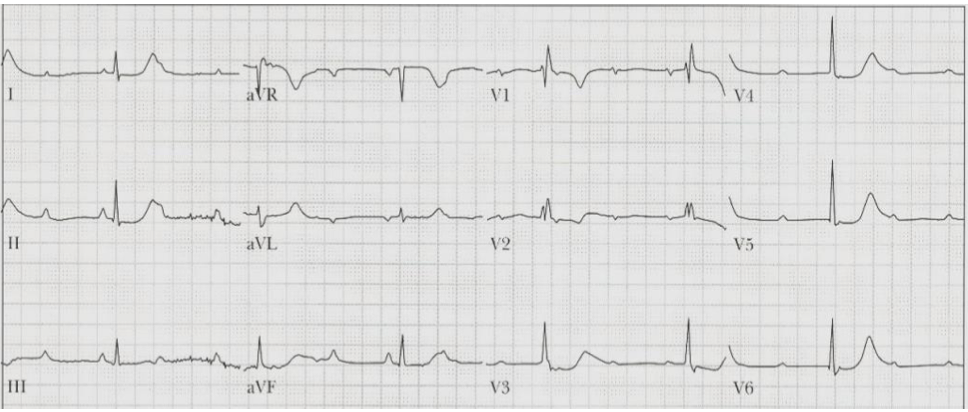
Sample Answer
Sinus tachycardia, complete heart block, junctional escape rhythm, RBBB, diffuse ischemia.
In this example, I would give you 1 point for identifying the atrial rhythm (ST), one point for identifying CHB, 1 point for identifying that there is a junctional escape, 1 point for identifying the RBBB, and one point for noting the ischemia (ST depression in multiple leads). In other words… be complete in your interpretation!
Be sure that you don't just describe what you see....interpret the ECG! For example, I don't want you to say "ST depression in __ leads." I want you to say "diffuse ischemia."
Also, in your interpretation, if you think there's an underlying issue, say it.
For example, if you think the underlying dx is hyperK, or TCA OD, or Brugada, etc., say it.
Don't assume that points correlate with the number of things I am looking for. For example, if something is worth 3 points, that does NOT mean there are 3 things you need to list! Points are allotted based on how important I think something is.
Also, for all of the questions below, please assume that a cath lab is not immediately available to you.
9. 56 yo W presents with chest pain. BP 89/55. What is the FULL ECG diagnosis? (2 points) Based on current U.S. (ACC) guidelines, identify which lead(s) is/are diagnostic for the condition you identified above. (2 points, but lose a point if you identify leads that are not diagnostic) 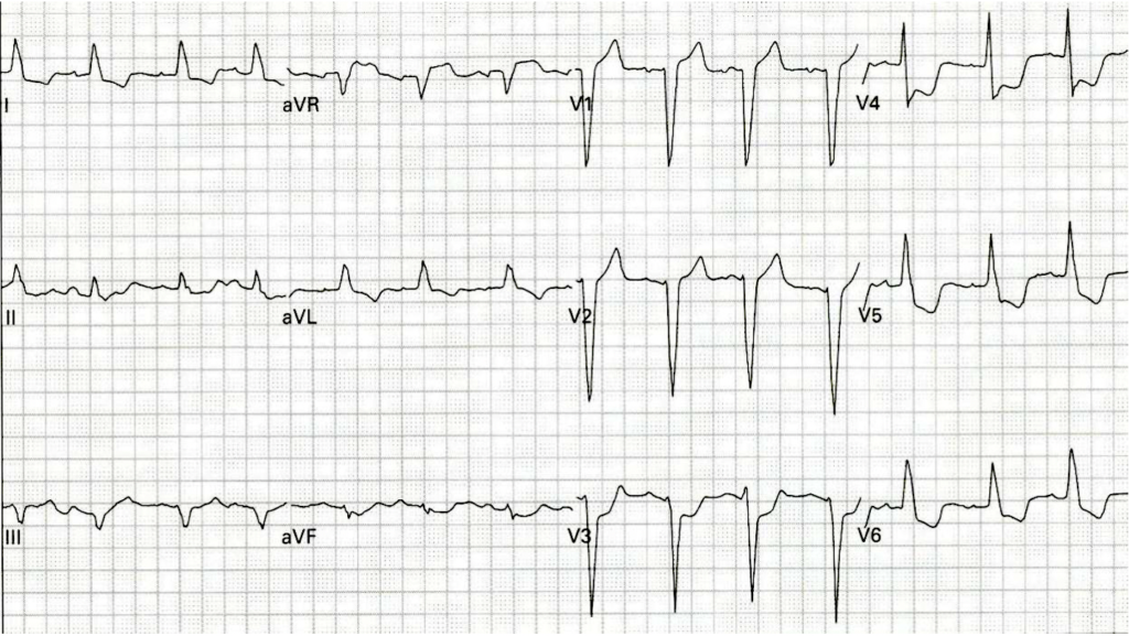
Question # 9 Answer
LBBB does not make ischemia uninterpretable
The first case this week involves a 56-year-old woman with chest pain and hypotension. The ECG shows sinus rhythm with left bundle branch block. Dr. Mattu reviews the ECG features supporting LBBB, including a wide QRS, deep broad S waves in V1-V3, leftward axis, and absence of Q waves in the lateral leads.
The critical diagnosis is not just LBBB. This patient has acute coronary occlusion in the presence of LBBB.
Dr. Mattu reviews the Sgarbossa framework:
- Sgarbossa A: Concordant ST elevation ≥1 mm in a lead with a positive QRS complex
- Sgarbossa B: Concordant ST depression ≥1 mm in V1, V2, or V3
- Modified Sgarbossa C: Excessively discordant ST elevation, usually defined as ST elevation more than 25% of the depth of the preceding S wave
In this case, V3 meets Sgarbossa B criteria because there is ST depression in V3 in the setting of LBBB. aVR also qualifies because the ST elevation is excessively discordant relative to the depth of the S wave, satisfying modified Sgarbossa criteria.
The leads Dr. Mattu wanted identified were V3 and aVR.
In LBBB, the direction and proportionality of ST deviation matter
Dr. Mattu reinforces the concept of appropriate discordance. In LBBB, the ST segment and T wave usually move opposite the main direction of the QRS complex. A predominantly positive QRS may have some ST depression; a predominantly negative QRS may have some ST elevation.
The problem is not discordance itself. The problem is:
- ST deviation in the same direction as the QRS when it should be discordant
- ST depression in V1-V3
- ST elevation that is excessively discordant relative to the preceding S wave
This proportional approach is why modified Sgarbossa is so useful. It avoids treating every discordant ST elevation in LBBB as abnormal while still identifying dangerous, excessive discordance.
10. A 69 yo W presents with dyspnea and vomiting. What is the diagnosis? (2 points) Following this ECG, the patient had a cardiac arrest and died. What did the autopsy show as the cause of death? Please be as specific as you can. (2 points) 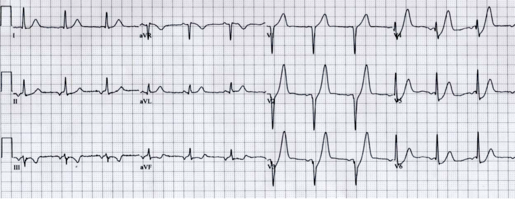
Question # 10 Answer
De Winter T waves are an LAD occlusion pattern and cath lab activation criterion
The second case involves a 69-year-old woman with dyspnea and vomiting. After the ECG, she arrests and dies. The question asks for the diagnosis and what autopsy would show as the cause of death.
The key ECG finding is upsloping ST depression in the mid-precordial leads, especially V2-V5, leading into large, tall, symmetric T waves. This is the De Winter T-wave pattern.
Dr. Mattu describes this as an ECG pattern associated with critical proximal LAD occlusion. It is now recognized in ACC guidance as a pattern that should prompt emergent cath lab activation.
The expected autopsy finding is therefore a critical proximal LAD occlusion or unstable proximal LAD blockage.
De Winter pattern is not "just ST depression"
A major trap is interpreting the De Winter pattern as nonspecific ST depression or subendocardial ischemia. The pattern is more specific than that.
The key morphology is:
- Upsloping ST depression at the J point
- Tall, symmetric, hyperacute-appearing T waves
- Most prominent in the mid-precordial leads
- Often representing proximal LAD occlusion physiology
In the right clinical context, this should be treated as a STEMI equivalent, not as a low-risk NSTEMI pattern.
11. Describe how lead V6 can be used to distinguish early repolarization from acute pericarditis and STEMI. Feel free to draw diagrams to help clarify your answer. (2 points)
Question # 11 Answer
Diffuse ST elevation has a focused but important differential
This question asks how lead V6 can help distinguish early repolarization from acute pericarditis and STEMI.
Dr. Mattu frames the differential for diffuse ST elevation as:
- Large STEMI
- Acute pericarditis
- Benign early repolarization
- Ventricular aneurysm
He notes that ventricular aneurysm is less commonly seen now than in the thrombolytic era, although it remains a board-exam favorite. In real ED practice, the more common practical differential is STEMI versus pericarditis versus early repolarization.
The ST/T ratio in V6 can help distinguish early repolarization from pericarditis and STEMI
The key measurement in lead V6 is the ratio of: ST-segment elevation height / T-wave height
Dr. Mattu explains:
- If the ST elevation is more than 25% of the T-wave height in V6, that favors acute pericarditis or STEMI
- If the ST elevation is less than 25% of the T-wave height in V6, that favors benign early repolarization
This is not a stand-alone rule that replaces clinical judgment, serial ECGs, reciprocal-change assessment, or overall pattern recognition. But it is a useful discriminator when early repolarization and pericarditis look similar.