A Closer Look at the J Point
ECG Weekly Workout with Dr. Amal Mattu
HPI
A 72-year-old man undergoes a prehospital 12-lead ECG. The tracing appears to show ST segment elevation in leads III, aVF, and aVR, raising concern for an inferior STEMI or high-risk ischemia. However, the ECG also demonstrates a markedly widened QRS complex with a right bundle branch block pattern.
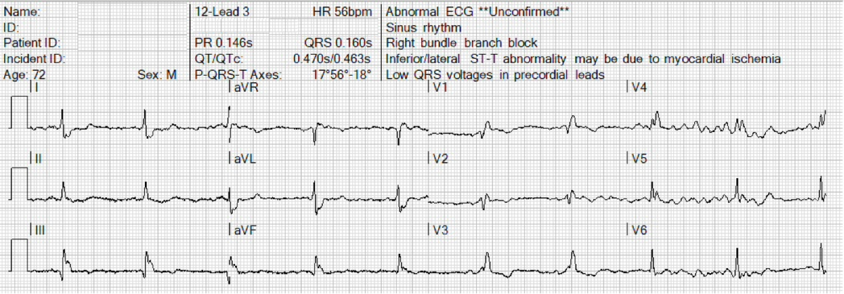
Before calling the cath lab, review the ECG carefully and consider:
-
- Is the apparent ST segment elevation real?
- How can a wide QRS complex create pseudo–ST segment elevation or depression?
- What is the most reliable way to locate the true J point when the end of the QRS is unclear?
