
HPI
This week we review the answers to the last 6 cases from the UMEM Residency ECG Competition. Make sure to attempt to answer the questions before clicking the red boxes to reveal answers and teaching pearls!
Last 6 Question Review Video:
Case 7. 56 yo M presents with lightheadedness and low BP. What is the likely cause of the low BP?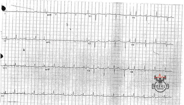
Large pericardial effusion or tamponade, suggested by tachycardia, low voltage QRS and electrical alternans
Low Voltage Definition
- Specific Definition
- QRS amplitudes in limb leads all < 5 mm OR in all chest leads < 10mm
- Sensitive Definition
- QRS amplitudes in I+II+III < 15 mm OR V1+V2+V3 < 30 mm
Low Voltage QRS Differential
- "Low Power"
- Myxedema (severe hypothyroidism)
- Infiltrative diseases (Amyloid, Sarcoid, etc.)
- End stage cardiomyopathy
- Conduction blockage
- Fluid/Effusion (pericardial or pleural)
- Fat (obesity)
- Air (COPD)
Pearls:
New low voltage ECG + Tachycardia = Pericardial effusion until proven otherwise! New low voltage ECG + Bradycardia = Myxedema until proven otherwise!
For more information about low voltage, review the case from April 6, 2015.
Case 8. A 36-year-old man presents weak and dizzy. What is the most important immediate treatment? Name 4 other causes of a large S-wave in lead I.
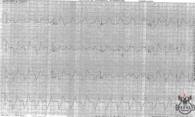
Calcium for Severe Hyperkalemia
ECG findings in Hyperkalemia
- Peaked T-waves
- Widening of the QRS (often marked)
- Prolonged PR-interval
- Flattening and eventual loss of P-waves
- Tachydysrrhythmias
- Advanced AV Blocks and sinus pauses
- Fascicular & Bundle Branch Blocks
- Pseudo ACS with ST-segment changes (can mimic STEMI)
- Pseudo Brugada syndrome pattern
- Sine wave morphology
Causes of Rightward Axis Deviation
- Hyperkalemia
- Na+ channel blocker toxicity
- Pulmonary HTN - Acute (PE) & chronic lung disease (COPD)
- Right ventricular hypertrophy
- Left posterior fascicular block
- Lateral MI (from Q-waves in lead I)
- Ventricular ectopy (VT)
- Lead misplacement / Dextrocardia
- Normal thin adults with horizontally positioned hearts
Pearl: When you see a wide QRS rhythm with a rightward axis, think of HYPERKALEMIA or other sodium channel blocker toxicities.
Bonus Pearl: Massive STE can mimic wide QRS complexes (typically only in a few leads, but usually not with a rightward axis!).
For more practice on this topic, review the case from Jan 12, 2015.
Case 9. A 36-year-old man presents with lightheadedness. What is the patient's rhythm?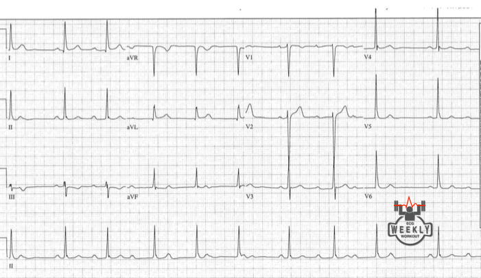
Mobitz I (Wenckeback)
Differential for Non-conducted P-waves (P:QRS > 1)
- Blocked Premature Atrial Complexes
- P-waves are irregular, the non-conducted P-waves come early
- Mobitz I (Wenckebach)
- P-waves are regular (P-P interval is constant)
- Progressive PR-interval lengthening before a non conducted beat
- Mobitz II
- P-waves are regular (P-P interval is constant)
- Some impulses fail to conduct to the ventricles WITHOUT progressive PR-interval lengthening
- AV Dissociation, 3rd degree AV Block
- P-waves are regular (P-P interval is constant)
- PR-interval is randomly changing
To learn more about "electrocardiographic polyuria" review the case from March 2, 2015.
Case 10. A 60-year-old woman presents with dyspnea. What is the cause of the patient's wide QRS? Name 5 other causes of a wide QRS.
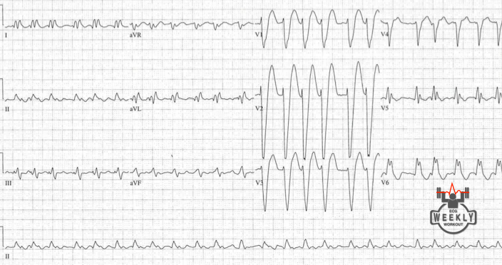
Nonspecific intraventricular conduction delay
Causes of QRS prolongation
- Metabolic/Electrolytes (acidosis, hyperkalemia)
- Medications - Na+ channel blocking toxicity (i.e. TCA's & Anti-arrhythmics)
- Ventricular Ectopy
- Bundle Branch Blocks and Paced rhythms
- Pre-excitation (WPW)
- Nonspecific intraventricular conduction delay (ex. from LVH or cardiomyopathy)
Pearl: LBBB should not have any Q-waves in lateral leads, even small Q-waves in the lateral leads obviates the diagnosis of LBBB.
Case 11. A 48-year-old man presents with fatigue. What is the rhythm?
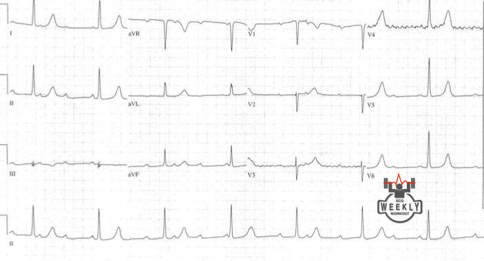
Complete heart block with junctional escape rhythm
To learn more about complete heart block, review the case from April 27, 2015.
Case 12. A 60-year-old woman presents with palpitations. What is your full ECG interpretation? Bonus: What is the "BIX rule?"
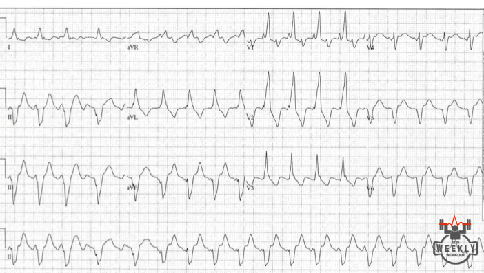
Atrial flutter with RBBB and Q-waves in inferior and lateral leads
Bonus: The Bix rule, described by cardiologist Harold Bix, states that if a P-wave is located halfway between two QRS complexes, there is a good chance that P-wave's are also buried inside the QRS complexes.
Reference:
Nikolić G. The Bix rule. Heart Lung 2008;37(4):321-2. PMID: 18620109.