
HPI
This week we review the answers to questions 12-14 from the 14th Annual UMEM Residency ECG Competition. Make sure to attempt to answer the questions yourself before watching the video or clicking the answer boxes in the summary section to reveal the answers and take home points!
Instructions from Dr. Mattu: In some of the following questions, I'll ask you for a FULL ECG interpretation/diagnosis. Here's what I am asking for:
SAMPLE QUESTION: A 58 yo M presents with chest pain and the ECG noted below. What is the FULL ECG diagnosis? (5 points)
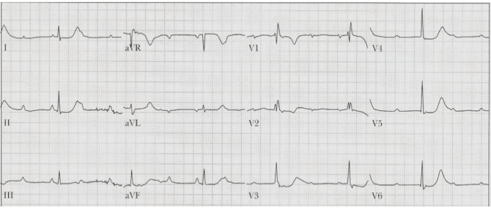
Sample Answer
Answer:Sinus tachycardia, complete heart block, junctional escape rhythm, RBBB, diffuse ischemia
In this example, I would give you 1 point for identifying the atrial rhythm (ST), one point for identifying CHB, 1 point for identifying that there is a junctional escape, 1 point for identifying the RBBB, and one point for noting the ischemia (ST depression in multiple leads). In other words...be complete in your interpretation!
Be sure that you don't just describe what you see....interpret the ECG! For example, I don't want you to say "ST depression in __ leads." I want you to say "diffuse ischemia."
Also, in your interpretation, if you think there's an underlying issue, say it. For example, if you think the underlying dx is hyperK, or TCA OD, or Brugada, etc., say it.
Don't assume that points correlate with the number of things I am looking for. For example, if something is worth 3 points, that does NOT mean there are 3 things you need to list! Points are allotted based on how important I think something is.
Also, for all of the questions below, please assume that a cath lab is not immediately available to you.
12. List the ECG criteria for the Aslanger pattern for acute coronary occlusion. (2 points)
Question # 12 Answer
Aslanger pattern identifies occlusion that does not meet STEMI criteria
Question 12 asks for the ECG criteria for the Aslanger pattern, a relatively recently described pattern of acute coronary occlusion. Dr. Mattu emphasizes that this pattern usually reflects acute occlusion involving either the left circumflex or right coronary artery.
The key danger is that the ECG may show ST elevation in only one inferior lead, usually lead III. Since the computer algorithm is generally programmed to look for ST elevation in two contiguous leads, it may not identify this as a STEMI. Clinicians can miss it for the same reason.
The Aslanger criteria reviewed in the episode are:
- ST elevation in lead III, but not in the other inferior leads
- ST elevation in V1 greater than V2, or V1 elevated while V2 is isoelectric
- Lateral ST depression, especially in V4-V6, with upright terminal T waves
This is an occlusion pattern, not a benign abnormality. It should prompt concern for an acute RCA or LCx occlusion even when classic STEMI criteria are not met.
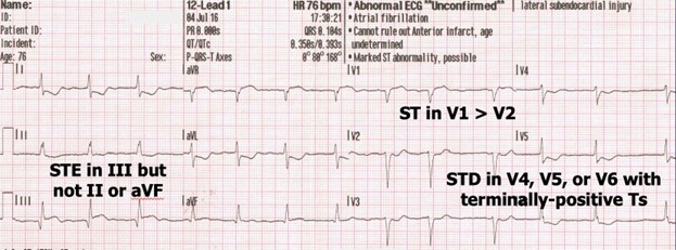
Aslanger pattern can fool both humans and computers
Dr. Mattu stresses that the Aslanger pattern is often missed because it violates the usual "two contiguous leads" STEMI framework. Lead III may be the only inferior lead with ST elevation, so both clinicians and computer interpretations may dismiss it.
The lateral leads are critical. The pattern is not simply "lead III elevation." It is the combination of isolated lead III ST elevation, V1/V2 relationship, and lateral ST depression with upright terminal T waves.
The practical takeaway: when a patient has ischemic symptoms and isolated ST elevation in lead III, do not stop there. Look carefully at V1, V2, and the lateral precordial leads.
13. Describe what "T wave alternans" is and why it is significant when found. (2 points)
Question # 13 Answer
T wave alternans is a warning sign for impending torsades
T wave alternans is beat-to-beat variation in the T wave. The T wave may alternate in size, shape, or magnitude from one beat to the next. It may be upright or inverted; the key feature is the alternating beat-to-beat change.
Dr. Mattu contrasts this with electrical alternans, where the QRS amplitude alternates and often suggests pericardial effusion. T wave alternans is different and much more concerning for electrical instability.
The major clinical significance is that T wave alternans is a strong warning sign for impending torsades de pointes.
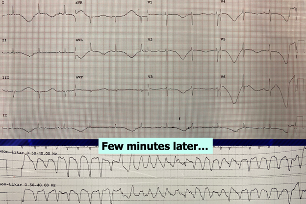
T wave alternans is more ominous than QT prolongation alone
A prolonged QT interval should always prompt concern, medication review, electrolyte evaluation, and correction of reversible causes. But Dr. Mattu emphasizes that T wave alternans is a more alarming short-term predictor of torsades than QT prolongation alone.
When T wave alternans is seen, the patient may deteriorate within minutes: take it seriously, correct electrolytes aggressively, consider empiric magnesium, and place defibrillator pads on the patient.
There is not strong trial evidence for an empiric protocol based solely on T-wave alternans, but clinically, this is a high-risk ECG finding that should trigger immediate preparation.
14. Describe the Barcelona criteria for diagnosing acute coronary occlusion in the presence of a LBBB. (2 points)
Question # 14 Answer
Barcelona criteria are another method for diagnosing OMI in LBBB
Dr. Mattu notes that in North America, clinicians more commonly use classic and modified Sgarbossa criteria. In parts of Europe, the Barcelona criteria are more discussed.
He is cautious about overpromoting them: the modified Sgarbossa criteria have more supporting evidence and are incorporated into national guidance, whereas the Barcelona criteria have less evidence and are not used as widely in North America.
Still, the Barcelona criteria are worth knowing.
Barcelona criteria overlap with Sgarbossa but broaden the ST-depression rule
Dr. Mattu reviews the modified Sgarbossa criteria first:
- Concordant ST elevation ≥1 mm in any lead
- Concordant ST depression ≥1 mm in V1-V3
- Excessively discordant ST elevation, defined proportionally as ST elevation at least 25% of the depth of the preceding S wave
The Barcelona criteria keep the first criterion the same:
- Concordant ST elevation ≥1 mm in any lead
But they broaden the second criterion:
- Concordant ST depression ≥1 mm in any lead, not just V1-V3
That is one major difference from traditional Sgarbossa.
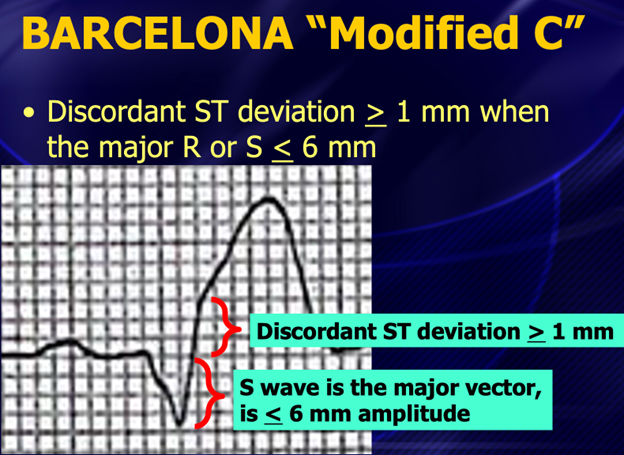
Barcelona criteria also use small QRS complexes with discordant ST deviation
The third Barcelona criterion is different from modified Sgarbossa.
Instead of using a proportional ST/S-wave ratio, the Barcelona criteria look for a small QRS complex with discordant ST deviation.
The rule Dr. Mattu teaches:
- If the QRS complex is ≤6 mm, then ST deviation ≥1 mm in the opposite direction is considered positive
This can apply to discordant ST elevation or discordant ST depression, as long as the total QRS amplitude is small enough.
The rationale is that even 1 mm of discordant ST deviation becomes suspicious when the QRS complex is small.
Video
Keyboard shortcuts
- , / .
- Slower / faster by 0.1×
- Shift + , / .
- Slower / faster by 0.25×
- /
- Back to 1×
- Space or K
- Play / pause
- ← / →
- Back / forward 10s
- Shift + ← / →
- Back / forward 5s
- M
- Mute
Your speed is remembered for every video.
Written Summary & Key Teaching Points
- Aslanger pattern is an occlusion MI pattern that may not meet traditional STEMI criteria.
- Do not rely on the computer to detect Aslanger pattern. It often has ST elevation in only lead III.
- Aslanger criteria include isolated ST elevation in III, V1 ST elevation greater than V2, and lateral ST depression with upright terminal T waves.
- Aslanger pattern predicts acute RCA or LCx occlusion.
- T-wave alternans is beat-to-beat variation in T-wave size, shape, or magnitude.
- T-wave alternans is a strong warning sign for impending torsades de pointes.
- T-wave alternans should trigger immediate risk mitigation: pads, electrolyte correction, medication review, and consideration of magnesium.
- Barcelona criteria are an alternative method for diagnosing occlusion MI in LBBB, but modified Sgarbossa has stronger evidence and broader North American adoption.
- Barcelona criteria broaden concordant ST depression to any lead, not only V1–V3.
- Barcelona criteria treat ≥1 mm discordant ST deviation as positive when the QRS complex is ≤6 mm.