
HPI
This week we review the answers to questions 1-4 from the 14th Annual UMEM Residency ECG Competition. Make sure to attempt to answer the questions yourself before watching the video or clicking the answer boxes in the summary section to reveal the answers and take home points!
Instructions from Dr. Mattu: In some of the following questions, I'll ask you for a FULL ECG interpretation/diagnosis. Here's what I am asking for:
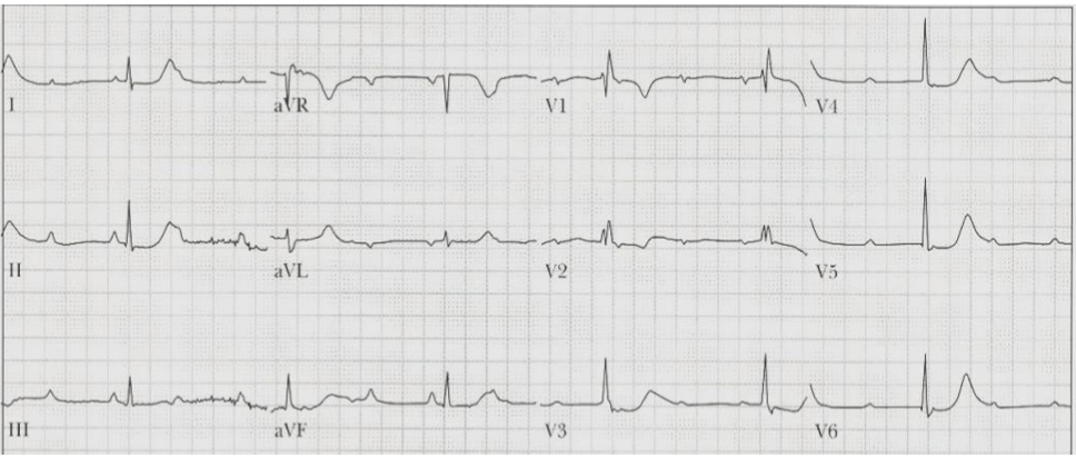
SAMPLE QUESTION: A 58 yo M presents with chest pain and the ECG noted below. What is the FULL ECG diagnosis? (5 points)
Sample Answer
Answer:
Sinus tachycardia, complete heart block, junctional escape rhythm, RBBB, diffuse ischemia
In this example, I would give you 1 point for identifying the atrial rhythm (ST), one point for identifying CHB, 1 point for identifying that there is a junctional escape, 1 point for identifying the RBBB, and one point for noting the ischemia (ST depression in multiple leads). In other words...be complete in your interpretation!
Be sure that you don't just describe what you see....interpret the ECG! For example, I don't want you to say "ST depression in __ leads." I want you to say "diffuse ischemia."
Also, in your interpretation, if you think there's an underlying issue, say it. For example, if you think the underlying dx is hyperK, or TCA OD, or Brugada, etc., say it.
Don't assume that points correlate with the number of things I am looking for. For example, if something is worth 3 points, that does NOT mean there are 3 things you need to list! Points are allotted based on how important I think something is.
Also, for all of the questions below, please assume that a cath lab is not immediately available to you.
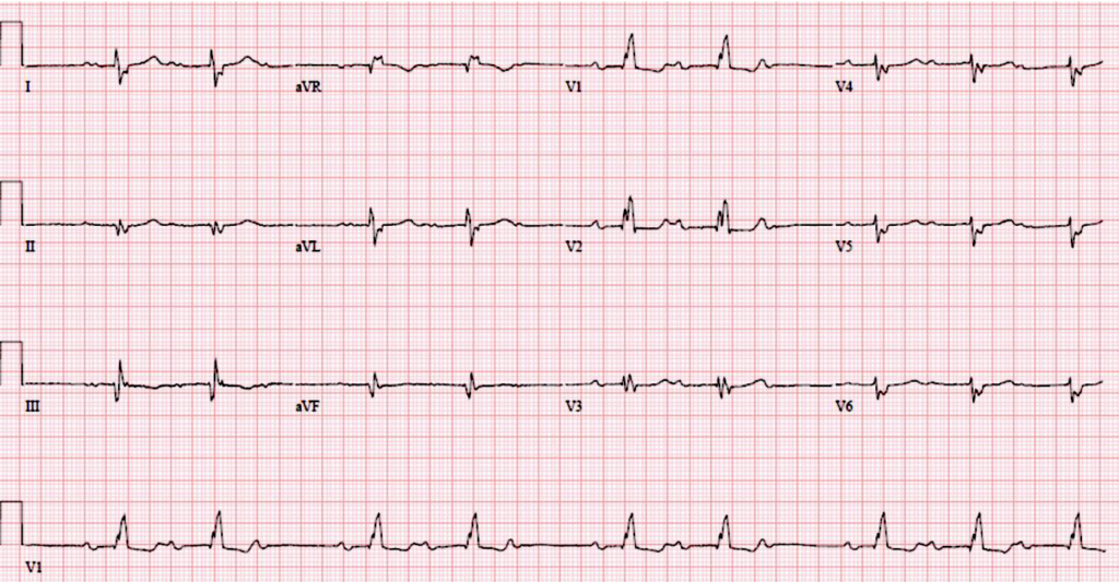
1. A 75 yo M presents after an episode of near-syncope. His BP is 110/65. He is awake and alert. What is the FULL ECG diagnosis? (4 points)
Question # 1 Answer
Grouped beating should trigger careful P-wave analysis
The first case involves a 75-year-old man with near syncope and grouped beats on ECG. Dr. Mattu points out that grouped beating often suggests Mobitz AV block, but frequent PACs can create a similar appearance.
The key maneuver is to march out the P waves. In this case, the P-P interval is fairly regular, arguing against PACs as the explanation. The PR interval progressively lengthens before a dropped QRS complex, confirming Mobitz I AV block.
Dr. Mattu also reinforces that this is not complete heart block, because complete heart block requires a regular escape rhythm. An irregular ventricular response argues against complete AV dissociation with a stable escape rhythm.
The full ECG interpretation included:
- Sinus rhythm
- Mobitz I AV block
- Right bundle branch block
- Evidence of prior inferior MI
The prior inferior MI is clinically relevant and should not be ignored. If a patient does not know they have evidence of a prior infarction, that finding may warrant follow-up.
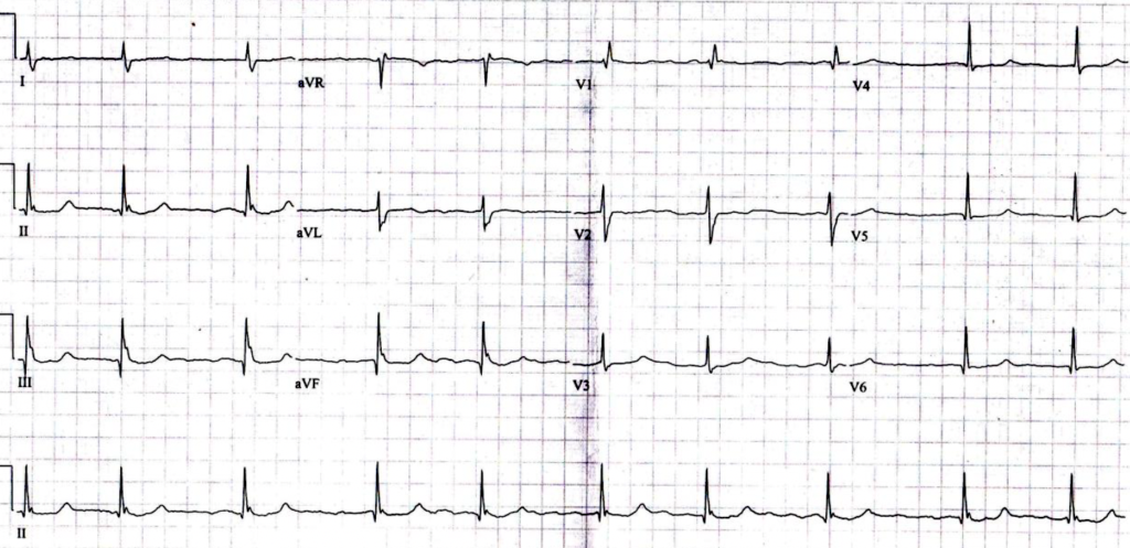
2. A 36 yo M presents with gradual progression of dyspnea on exertion, palpitations, and lower extremity (LE) edema over the past 2-3 weeks. He has a long history of exercise intolerance and intermittent atrial fibrillation. What diagnosis is suggested by the ECG as the underlying cause of his symptoms? (2 points) What do you expect to find on his exam (aside from JVD & LE edema)? (2 points)
Question # 2 Answer
Atrial septal defect can produce right heart strain findings on ECG
The second case involves a 36-year-old man with gradual dyspnea on exertion, palpitations, lower extremity edema, long-standing exercise intolerance, and intermittent atrial fibrillation. The question asks for the underlying diagnosis suggested by the ECG and expected exam findings.
The ECG shows atrial fibrillation, but that is already known. More important are the subtle findings suggesting right heart strain, including:
- S wave in lead I
- Incomplete right bundle branch block pattern
- Tall R wave in V1
- Inferior-lead notching of the QRS complex
Dr. Mattu identifies the inferior-lead notching as the crochetage pattern, which is classically associated with secundum atrial septal defect, especially when seen in all three inferior leads.
The underlying physiology is a left-to-right shunt, causing chronic right-sided volume overload. This produces right heart strain and eventually symptoms such as dyspnea, exercise intolerance, edema, palpitations, and atrial arrhythmias.
Expected exam findings include:
- Systolic ejection murmur at the left upper sternal border
- Fixed split S2
Additional findings may include a diastolic rumble and a hyperdynamic precordium. Dr. Mattu makes the point that physical examination still matters, even in the era of ultrasound.
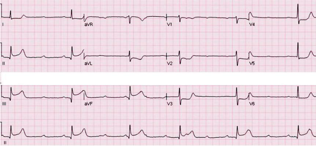
3. A 56 yo W presents w/chest tightness and dyspnea. BP is 125/75. What is the FULL ECG diagnosis? (4 points)
Question # 3 Answer
High-grade AV block is not the same as complete heart block
The third case involves a 56-year-old woman with chest tightness and shortness of breath. The ECG shows many nonconducted P waves, but Dr. Mattu cautions against immediately labeling the rhythm as complete heart block.
The distinction depends on the ventricular rhythm. In complete heart block, the escape rhythm should be regular. In this case, the QRS complexes are irregular, and some ventricular beats occur earlier than expected. This suggests that some atrial impulses are intermittently conducting and capturing the ventricles.
The correct rhythm diagnosis is therefore high-grade AV block, or AV dissociation without complete heart block, rather than complete heart block.
The ECG also shows:
- Inferior STEMI
- Posterior extension, suggested by horizontal ST depression with upright T waves in the anterior leads
- Sinus rhythm or sinus tachycardia as the underlying atrial rhythm
- Likely junctional escape rhythm with intermittent capture beats
The key lesson is that complete heart block requires a regular escape rhythm. If the QRS complexes are irregular and some beats occur early due to capture, the better diagnosis is high-grade AV block.
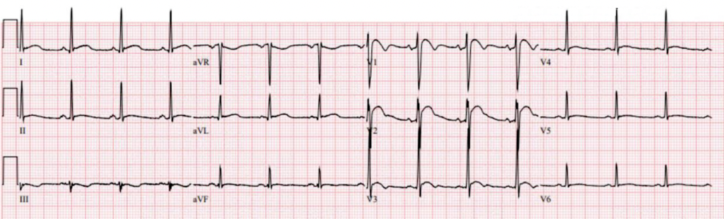
4. A 35 yo M presents with vomiting and weakness. His BP is 105/58. What is the diagnosis? (2 points) What is the most important therapy for this patient in the first hour? (2 points)
Question # 4 Answer
Hypercalcemia can mimic anterior STEMI by shortening the ST segment
The fourth case involves a 35-year-old man with vomiting, weakness, and borderline hypotension. At first glance, the ECG may appear concerning for anterior STEMI. Dr. Mattu uses this case to reinforce the importance of looking at the entire ECG before anchoring.
The rhythm, axis, PR interval, and QRS duration are relatively unremarkable. The key abnormality is a markedly short QT interval.
A short QT interval should suggest:
- Hypercalcemia
- Digoxin effect
- Rare congenital short QT syndrome
In this adult patient with vomiting and weakness, the correct diagnosis is severe hypercalcemia. The calcium level was reportedly around 17.9 mg/dL, likely related to malignancy.
Hypercalcemia shortens the ST segment. As the ST segment shortens or disappears, the T wave appears "smooshed" against the QRS complex, creating an apparent ST elevation pattern, especially in the anterior leads. This can mimic anterior STEMI.
Clues against STEMI include:
- Very short QT interval
- Lack of reciprocal ST depression
- Apparent ST elevation caused by loss of the ST segment rather than true ischemic STE
The most important early therapy is IV fluids. Dr. Mattu emphasizes that patients with severe hypercalcemia are typically volume depleted, and aggressive fluid resuscitation is the priority in the first hour.
Video
Keyboard shortcuts
- , / .
- Slower / faster by 0.1×
- Shift + , / .
- Slower / faster by 0.25×
- /
- Back to 1×
- Space or K
- Play / pause
- ← / →
- Back / forward 10s
- Shift + ← / →
- Back / forward 5s
- M
- Mute
Your speed is remembered for every video.
Written Summary & Key Teaching Points
- Grouped beating is not automatically Mobitz block. March out the P waves to distinguish Mobitz AV block from frequent PACs.
- Complete heart block should have a regular escape rhythm. An irregular QRS rhythm with intermittent capture beats is better described as high-grade AV block or AV dissociation without complete heart block.
- Do not ignore prior MI findings. Evidence of prior inferior MI is part of the ECG interpretation and may have important follow-up implications.
- Crochetage pattern in the inferior leads suggests secundum ASD. In the right clinical context, inferior QRS notching plus right heart strain findings should raise concern for atrial septal defect.
- Hypercalcemia can mimic anterior STEMI. Severe hypercalcemia shortens or abolishes the ST segment, making the T wave appear fused to the QRS complex.
- Short QT should trigger a differential. Think hypercalcemia, digoxin effect, and rare short QT syndrome.
- Severe hypercalcemia needs fluids early. IV fluids are the most important initial therapy in the first hour.