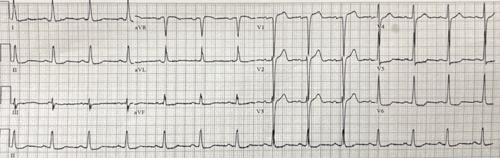
LVH with Strain Pattern Mimicking Anterior STEMI. Marked QRS voltage with deep S waves in V1–V3 and tall R waves in the lateral leads, accompanied by secondary repolarization abnormalities. Note the discordant ST elevation in the anterior leads with asymmetric ST–T changes, a classic LVH strain pattern that can closely resemble anterior occlusion but is typically stable and proportional to QRS amplitude.
