
HPI
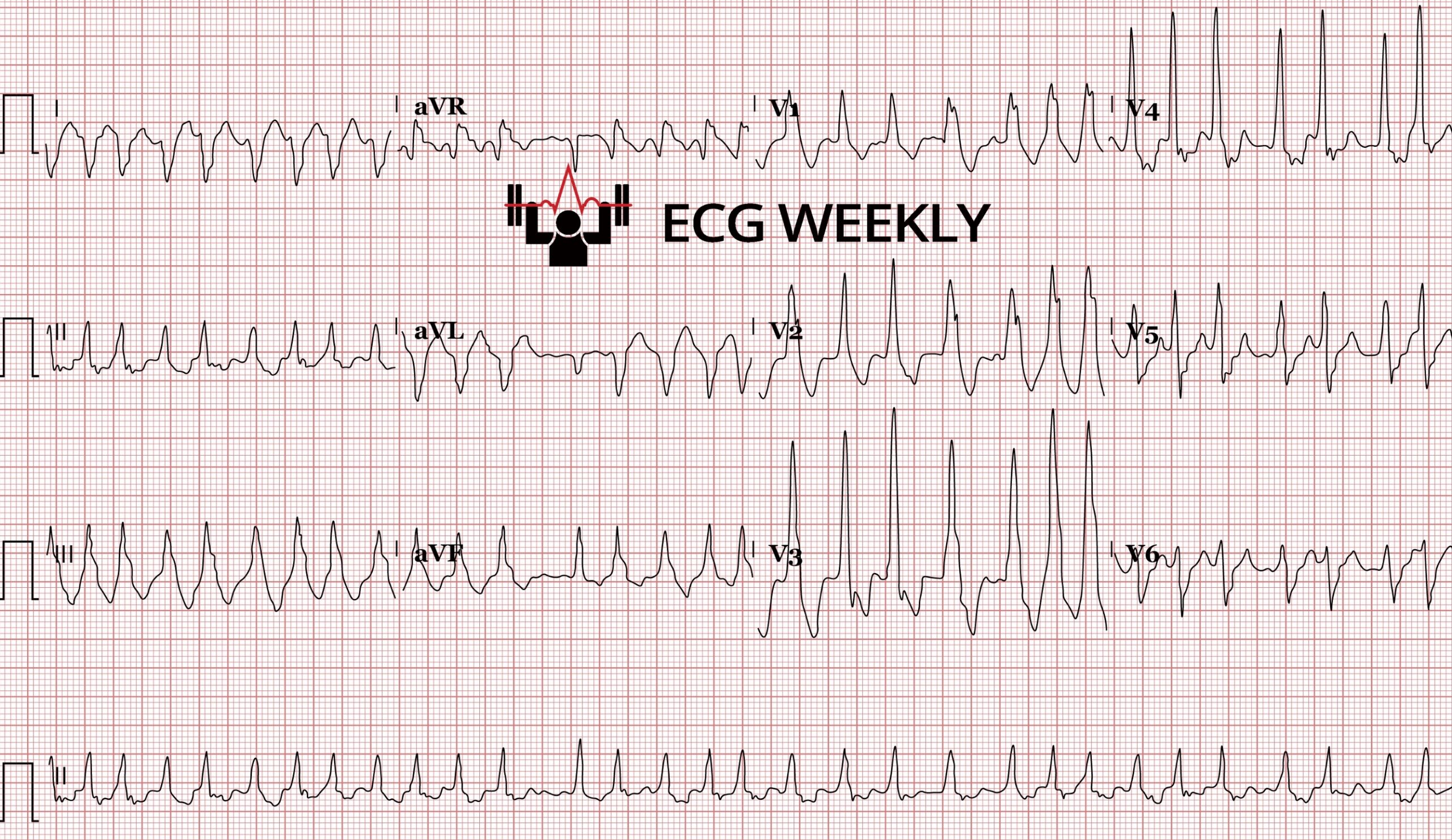
A 68-year-old man with PMHx of hypertension and tobacco use arrives at the emergency department with severe central chest pain and mild shortness of breath over the past 1-2 hours. The following initial ECG was obtained on arrival and interpreted as “normal”, a repeat ECG obtained 45 minutes later evolves into a large anterior STEMI:
Before watching this week’s workout, closely examine the ECG and ask yourself:
-
- Would you agree that the initial ECG was correctly interpreted as normal?
- Do you see any U-waves or terminal T-wave inversions?
- Is this ECG predictive of ischemia to any specific coronary artery distribution or can it predict a serious imminent infarction?
Video
Keyboard shortcuts
- , / .
- Slower / faster by 0.1×
- Shift + , / .
- Slower / faster by 0.25×
- /
- Back to 1×
- Space or K
- Play / pause
- ← / →
- Back / forward 10s
- Shift + ← / →
- Back / forward 5s
- M
- Mute
Your speed is remembered for every video.
Kudos
Written Summary & Key Teaching Points
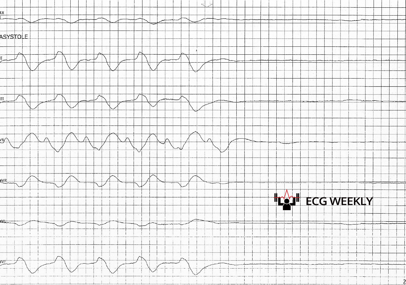 Case #10: Rapidly Progressive Hyperkalemia (Oct 26, 2020)
- Key Lesson: Hyperkalemia can escalate from mild ECG changes (peaked T-waves) to a sine wave pattern and asystole within minutes.
- Take-Home Point: Treat severe hyperkalemia immediately and aggressively—delays can be fatal.
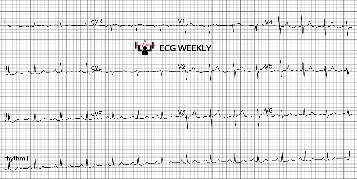 Case #9: Inverted U Waves Predict Impending Anterior MI (Jan 16, 2023)
- Key Lesson: Subtle inverted U waves in the antero-lateral leads can herald an imminent anterior myocardial infarction.
- Take-Home Point: Don’t ignore subtle U-wave changes; they can indicate serious, impending ischemia. Inverted U waves are not normal and very predictive of ischemic heart disease, can predict LAD or LMCA disease during pain or painless state.
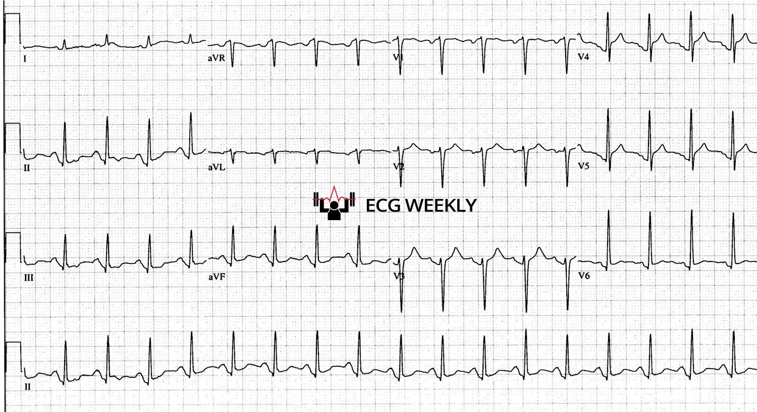 Case #8: Misdiagnosed ACS as Pericarditis (May 24, 2021)
- Key Lesson: Profound PR depression can occur in ischemia as well as pericarditis. Using the TP segment (not PR) for baseline can clarify ST segment changes.
- Take-Home Point: Don’t rely solely on PR depression for diagnosing pericarditis. Assess the ST segments carefully using the TP baseline to distinguish ACS from pericarditis (Note: TP segment is not reliable if the patient is very tachycardic).
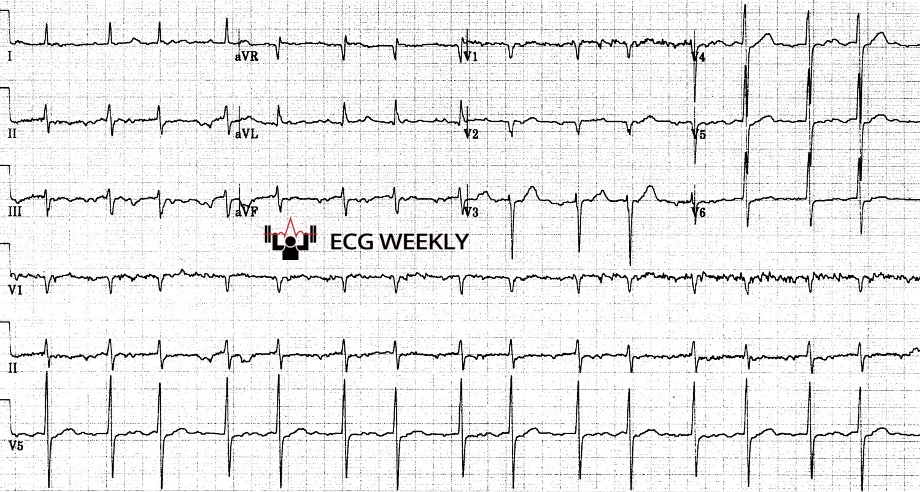 Case #7: Mobitz I Mistaken for Atrial Fibrillation (Dec 16, 2019)
- Key Lesson: A regularly irregular rhythm (grouped beats) can mimic AFib but may actually be Mobitz I (Wenckebach). Misdiagnosis can lead to unnecessary anticoagulation.
- Take-Home Point: Confirm true irregular irregularity before labeling a rhythm as AFib. Look for P waves and grouped beats to identify AV block patterns.
 Case #6: Defibrillator Synchronization Causing R-on-T VF (May 20, 2024 & Feb 6, 2022)
- Key Lesson: Before cardioversion, ensure the defibrillator syncs with the QRS and not the T wave. Shocking on the T wave can precipitate VF.
- Take-Home Point: Always verify that the machine’s sync markers align with the QRS complexes, not the T waves, to avoid an R-on-T phenomenon.
- Small, subtle ECG clues can predict catastrophic events.
- Always confirm your assumptions (e.g., AFib vs. Mobitz I, pericarditis vs. ischemia).
- Technology (like automatic sync mode) can mislead if not carefully checked.
- The importance of continuous learning: rare cases teach vital lessons that can save lives.