Unrecognized Killers in Emergency Electrocardiography
ECG Weekly Workout with Dr. Amal Mattu
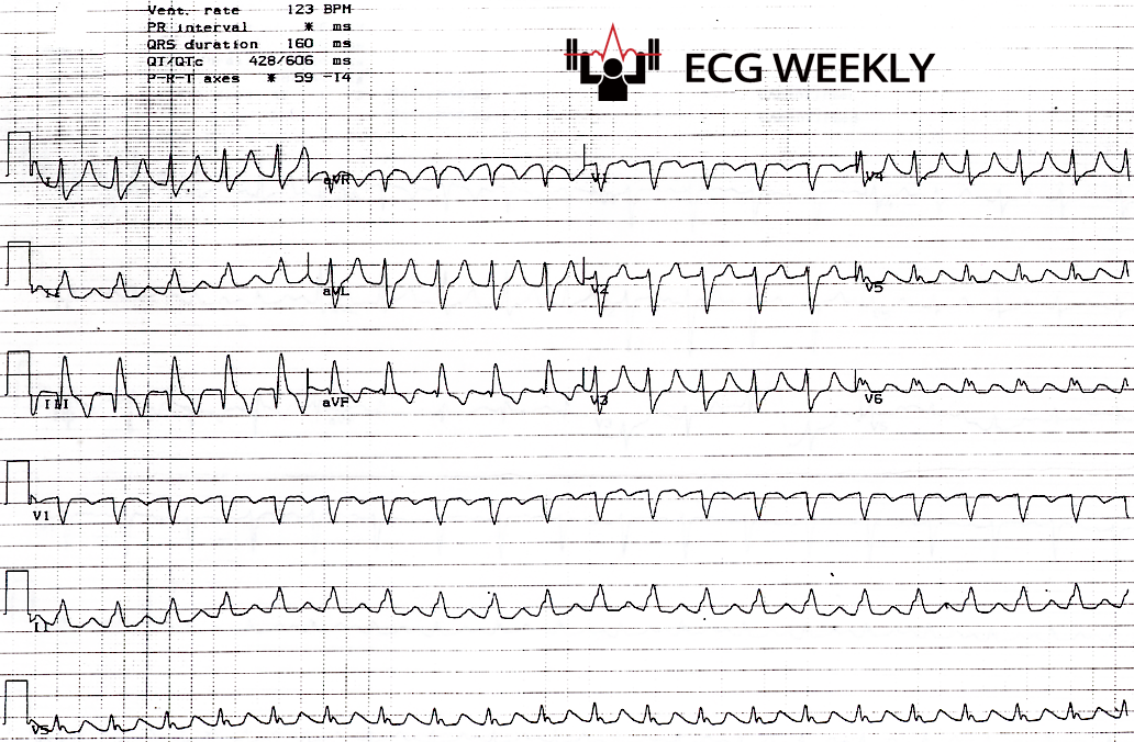
HPI
A 42-year-old man presents with fever, cough, shortness of breath, and vomiting. He appears toxic and dehydrated on arrival with suspected severe sepsis from multilobar pneumonia. He is later intubated for hypoxemic respiratory failure, after which there is a change in rhythm noted on the monitor. The following ECG is obtained which shows a regular wide complex tachycardia. He is treated with lidocaine for suspected stable ventricular tachycardia (VT), but develops asystole after lidocaine bolus and is not able to be resuscitated.
Before watching this week’s workout, carefully review the ECG and consider the following:
-
- Is this wide complex tachycardia VT?
- How would you manage this rhythm?
- What is the most likely cause of asystole and arrest after lidocaine bolus?
