
Rhythm Interpretation
Latest
UMMC Cases Part IV: Two Easily Missed ECG Diagnoses
A 73-year-old woman presents to the ED with palpitations and shortness of breath. Her ECG shows a regular narrow-complex tachycardia, and the computer interpretation is accepted by both the emergency…
UMMC Cases Part II: Grouped Beats, Slow VT, and Diffuse ST Elevation
A 63-year-old man presents with shortness of breath. His ECG shows an unusual rhythm with repetitive groups of two beats separated by pauses.
UMMC Cases Part I: Three ECGs That Reward a Second Look
A 62-year-old man presents with chest pain during a busy emergency department shift. His triage ECG is interpreted by the computer as showing ST-segment depression and possible ischemia. The tracing…
ECG Hallucinations: The Rhythms You See and the Rhythms You Miss
A patient presents with a regular narrow-complex tachycardia. The computerized interpretation identifies sinus tachycardia, and the rhythm appears superficially compatible with that diagnosis when lead II is reviewed. However, closer…
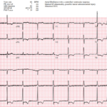
ECG Hallucinations: When Irregular Is Not Atrial Fibrillation
A 69-year-old man presents with chest pain. His ECG is interpreted by the computer as atrial fibrillation, a diagnosis that could significantly alter his evaluation and long-term management. The rhythm…
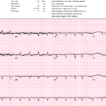
The Rhythm Behind the Irregularity
A 69-year-old woman presenting with sepsis gets the following ECG for tachycardia while febrile and shivering. The baseline is poor, atrial activity is difficult to identify, and the computer interpretation…
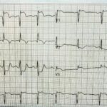
Three High Risk ECGs and the Right Next Move
A 34 year old man presents with acute chest pain radiating to the left arm associated with diaphoresis. He has hyperlipidemia and a family history of early coronary disease. The…
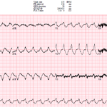
UMEM Cases, Part 4: When the Computer Misses the Rhythm and Flutter Fakes a STEMI
A 44-year-old man with severe cardiomyopathy, an LVAD, chronic amiodarone therapy, and an AICD presents with palpitations. His ECG shows a regular wide-complex tachycardia, but the rate is only 135….
UMEM Cases, Part 3: When the Diagnosis Seems Clear and When It Is Not
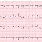
A 71-year-old man presents with shortness of breath, and his ECG is initially read as a junctional rhythm. On later review, it is even mistaken for atrial fibrillation. But the…
UMEM Cases, Part 2: When the ECG Conceals and When It Reveals
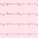
An 81-year-old woman presents with lightheadedness and marked bradycardia. Her ECG shows more P waves than QRS complexes, but the mechanism is not immediately clear. The key question is whether…
The Syncope ECG With Too Much P
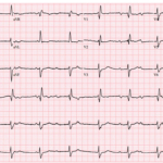
A 68-year-old man has syncope, then has a second syncopal episode while lying still on a stretcher during evaluation at an outpatient clinic. He is sent emergently to the ED….
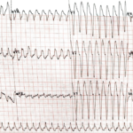
Preexcitation Pitfalls (Part 3): Wide, Irregular, Fast…Avoid AV Nodal Blockers
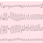
A 53-year-old man presents with palpitations and lightheadedness. The following ECG is obtained on arrival and appears very rapid and irregular with changing QRS morphologies. He starts showing signs of…
Preexcitation Pitfalls (Part 2): Wide, Regular, Fast…Treat It Like VT
A young man with recurrent palpitations presents to the emergency department hemodynamically stable during an episode. The arrival ECG shows a wide complex, regular tachycardia and the computer interpretation calls…
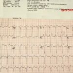
Preexcitation Pitfalls (Part 1): The “Inferior STEMI” That Isn’t
A critically ill 38-year-old man presents hypotensive, pale, and diaphoretic with abdominal pain and rectal bleeding. Upright chest X-ray shows free air under the diaphragm, and the patient is headed…
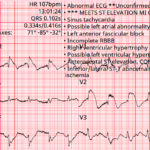
Baltimore City EMS ECGs: Pitfalls and Mimics (Part 2)
A 54-year-old man presents to the emergency department by EMS with acute shortness of breath. A prehospital ECG triggers a STEMI alert based on the computer interpretation. The tracing shows…
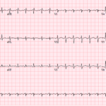
Three More ECG Pitfalls That Punish Anchoring Bias
A 51-year-old man with lung cancer presents with shortness of breath and tachycardia. The arrival ECG shows an S1Q3 pattern and seems to support a familiar diagnosis that would normally…
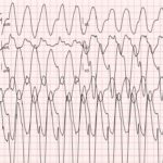
The Post-Arrest Wide Complex Rhythm That Was Not VT
A 35-year-old woman arrives at the emergency department after a witnessed seizure and brief cardiac arrest with ROSC. Her arrival ECG shows a very wide complex rhythm that the machine…
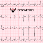
Recent ECG Case Highlights from the University of Maryland (Part III)
A 73-year-old man presents to the emergency department with nausea and vomiting. The following ECG was obtained on arrival and interpreted by the computer as “sinus tachycardia”, but something important…
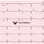
Recent ECG Case Highlights from the University of Maryland (Part II)
A 67-year-old man arrives at the emergency department complaining of lightheadedness. The following ECG is obtained and shows a slow, regular rhythm around 40 beats per minute. The P waves…
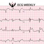
Recent ECG Case Highlights from the University of Maryland (Part I)
A 64-year-old man is brought to the ED by EMS just after achieving return of spontaneous circulation following cardiac arrest. He received chest compressions, epinephrine, and was defibrillated prior to…
Free Content
Jump on our email list for free tips and insights delivered to your inbox monthly. No spam - just quick pearls and ECG education.
Categories
- No categories

© 2026 ECG Weekly. All rights reserved. | Terms of Use | Privacy Policy
Powered by the member(dev) platform
 Loading...