
OMI Pattern
Results:

Occlusion MI: STEMI & Beyond Hub
Key Points: The ECG’s Primary Role in ACS Is Detecting Acute Coronary Occlusion: Acute coronary occlusion is a time-critical diagnosis in which rapid recognition and reperfusion can preserve myocardium and…
ST Depression V1-V4: OMI Pattern
Key Points: Posterior OMI can hide on the standard 12-lead. Acute posterior coronary occlusion may produce no diagnostic ST elevation in the conventional leads. Instead, posterior ST elevation is seen…
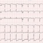
Four ECGs Hiding a High-Risk Coronary Pattern
A 56-year-old man presents with 1 week of concerning chest pain and shortness of breath. His ECG does not meet conventional STEMI criteria, but it shows subtle ST-segment abnormalities in…
Aslanger Pattern: OMI Pattern
Key Points: Aslanger pattern is an OMI pattern that can identify acute inferior occlusion despite not meeting traditional STEMI criteria. The key finding is ST segment elevation isolated to lead…
New RBBB + LAFB (Bifascicular Block) in ACS: OMI Pattern
Key Points: In a patient with ischemic symptoms, new RBBB + LAFB should raise concern for proximal LAD or septal ischemia until proven otherwise, especially if the patient has ongoing…
ST Elevation in aVR with Diffuse ST Segment Depression: OMI Pattern
Key Points: Pattern, not a STEMI equivalent. ST elevation in aVR (≥1 mm), often with ST elevation in V1 and diffuse ST depression (≥1 mm in ≥6 leads), represents high-risk…
South African Flag Sign: OMI Pattern
Key Points: Pattern: The South African Flag sign is the combination of ST elevation in I, aVL, and V2 with reciprocal ST depression in III. It is a subtle but…
STEMI (-) Occlusion MI: OMI Patterns
Key Points: Traditional STEMI criteria miss many acute coronary occlusions. A substantial proportion of true OMIs do not meet classic millimetric STEMI thresholds. OMI is a pathophysiologic diagnosis, not an…
Earliest Evidence of Occlusion MI
Key Points: ACS is dynamic. Coronary arteries can occlude, partially reperfuse, and re-occlude over minutes to hours, and the ECG can show these shifts before biomarkers do. The earliest actionable…
Early Reciprocal Changes: OMI Pattern
Key Points: STEMI criteria alone miss some acute coronary occlusions, so look for subtle “occlusion clues,” not just traditional STEMI criteria cutoffs. Minor ST elevation under 1 mm paired with…
Hyperacute T Waves: OMI Pattern
Key Points: Complexity of ACS: Arteries can spontaneously reperfuse or re-occlude. Understanding early ECG patterns of early acute ischemia can help detect dynamic changes and prompt rapid interventions. ECG Pattern…
Terminal QRS Distortion: OMI Pattern
Key Points: What it is: In V2 or V3, there is no S wave (the R does not descend below the PQ baseline) and no J wave (no notch/slur at…
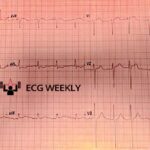
Aslanger’s Pattern and the Limits of Traditional STEMI Criteria
A 76-year-old man presents to the emergency department with shortness of breath, severe weakness, diaphoresis, and confusion. The following ECG is obtained on arrival:
Precordial Swirl: OMI Pattern
Key Points Definition: The precordial swirl sign refers to a rotational pattern of ST-segment abnormalities across the precordial leads, suggesting a dynamic and evolving occlusion myocardial infarction (OMI). It is…
Subtle ST Segment Elevation: OMI Pattern
Key Points: ACS Dynamics: Occluded arteries in ACS can spontaneously reperfuse and reocclude, making ECG findings variable. Limitations of Computer Interpretation: Subtle STE is often missed by computerized ECG interpretation;…
ECG Tags
- 5th Universal Definition of MI
- A-Z
- ACS Mimics
- ACS-OMI
- Activation
- Acute MI
- Advanced Level Curriculum
- Annual ECG Competition
- Anterior OMI
- Anterior STEMI
- Approach
- Arrest
- Arrhythmia
- Arrhythmogenic Cardiomyopathy
- Artifact
- ARVC
- ARVD
- Atrial Parasystole
- Attending
- AV Block
- aVF
- aVL
- aVR
- Axis
- Barcelona
- Basics
- Bix Rule
- Board Review
- Bradyarrhythmia
- Cath Lab
- Chest Pain
- Clumped Beats
- Competition
- Conduction
- Core
- Core Level Curriculum
- Critical ECG Patterns
- Curriculum
- DDx
- Delta
- Devices
- Diagonal Branch Occlusion
- Differential Diagnoses
- Diffuse ST Elevation
- Documentation
- Early Repolarization
- ECG Interpretation
- ECG Localization
- ECG Variant
- Education/Teaching
- Electrolytes
- Emergencies
- Emergent Cath Lab Activation
- EMS
- Expert Level Curriculum
- Flutter
- Flutter Waves
- Foundations Level Curriculum
- Guidelines
- High-Lateral STEMI
- Hub
- Hyperacute T waves
- Hypercalcemia
- Hyperkalemia
- Hypermagnesemia
- Hypocalcemia
- Hypokalemia
- Hypomagnesemia
- Hyponatremia
- Hypothermia
- I
- II
- Index
- Inferior OMI
- Inferior STEMI
- Intervals
- Irregular
- Ischemia
- Ischemia & Infarction
- J point
- J Waves
- JT
- Junctional Rhythm
- Juvenile T wave
- LAD Occlusion
- Lateral STEMI
- LBBB
- LCx Occlusion
- Lead Placement
- Left Atrial Enlargement (LAE)
- Life Savers
- LV an
- LV Aneurysm
- LVAD
- Mastery Level Curriculum
- Metabolic
- MI
- Mimics
- Morphology
- Narrow QRS
- Occlusion MI
- OMI Pattern
- Orthodromic AVRT
- Osborn Waves
- P Wave
- Paced Rhythms
- Pacemaker/ICD
- Paramedics
- Pauses
- PE
- Pediatrics
- Pericardial
- Pericarditis
- PGY-1
- PGY-2
- PGY-3
- PGY-4
- Post-Arrest
- post-arrest STEMI
- Post-Cardiac Arrest
- Posterior Extension
- Posterior MI
- Posterior STEMI
- potpourri
- PR
- Preexcitation
- Premature Complexes
- Prolonged QT
- Pulmonary Embolism
- Pulse-Tapping Artifact
- PVCs
- Q Wave
- QRS
- QT
- R Waves
- RAD
- Rate
- RBBB
- RCA Occlusion
- Regular
- Reperfusion
- Rhythm
- Right Atrial Enlargement (RAE)
- RR
- S Waves
- S1Q3T3
- Segments
- Seizure
- Serial ECGs
- Sgarbossa
- Shock
- South African Flag Sign
- ST
- ST Depression
- ST Elevation
- STEMI
- STEMI Equivalent
- STEMI Mimics
- STEMI Negative OMI
- Stepwise
- Stroke
- Structural
- Students
- SVT
- Syncope
- T Wave Inversion
- T Waves
- Tachyarrhythmia
- Tachycardia
- Tamponade
- Terminal QRS Distortion
- Toxicology
- TP
- Traditional STEMI Criteria
- U Waves
- Unstable
- V1-V4
- V2
- V5
- V6
- Vectors
- Ventricular Repolarization
- Ventricular Rhythms
- Ventricular Tachycardia
- Voltage
- Waveforms
- Wide QRS
- Workflow
- WPW
© 2026 ECG Weekly. All rights reserved. | Terms of Use | Privacy Policy
Powered by the member(dev) platform
 Loading...