Wide QRS
Results:

Appropriate Discordance
Key Points: Appropriate discordance refers to the expected secondary ST segment and T wave pattern seen with abnormal ventricular depolarization, especially LBBB and ventricular-paced rhythm. The ST segment and T…
WPW with Antidromic SVT (Antidromic AVRT)
Key Points: Antidromic AVRT is an AV re-entrant tachycardia that conducts antegrade down the accessory pathway and returns retrograde through the AV node (or another pathway), producing a regular wide-complex…
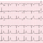
Bidirectional Ventricular Tachycardia (BiVT)
Key Points: BiVT is a regular wide-complex tachycardia with strict beat-to-beat alternation of QRS axis and/or bundle-branch pattern (often an approximately 180° frontal-plane axis flip). In adults, assume digoxin toxicity…
Four ECG Pitfalls That Punish Anchoring Bias
A 43-year-old woman with sharp left-sided chest pain and minimal cardiac risk factors has an initial ECG that is not diagnostic for STEMI. She looks stable, but one feature on…
Ventricular Tachycardia (VT): Core Overview
Key Points: VT is a ventricular-origin rhythm: ≥3 consecutive ventricular beats, QRS >120 ms, rate usually 120–250 bpm. Types include monomorphic VT, polymorphic VT, torsades (PMVT with long QT), ventricular…
Ventricular Tachycardia (VT) Mimics
Key Points: Initial Assumption: Any wide (QRS >120 ms), regular tachycardia should be considered ventricular tachycardia (VT) until clearly proven otherwise. VT Characteristics: VT generally has a ventricular rate of…
Pulseless Ventricular Tachycardia (pVT) Arrest
Key Points: Defibrillation First, Minimal Pauses: pVT is rapidly fatal without immediate shocks and high‑quality CPR. Charge defibrillator during compressions and resume compressions immediately after each shock. pVT is a…
Ventricular Fibrillation (VF) Arrest
Key Points: Defibrillation First, Minimal Pauses: VF is rapidly fatal without immediate shocks and high‑quality CPR. Charge during compressions and resume compressions immediately after each shock. Chaotic Electrical Activity: VF…
Ventricular Flutter (V-Flutter)
Key Points: Definition: A malignant ventricular tachyarrhythmia with a regular, sine-wave–like waveform at ~250–350 bpm, no isoelectric baseline, and no discernible P/QRS/T distinction. Clinical importance: Rapidly degenerates into ventricular fibrillation…
Unstable Tachyarrhythmias
Key Points: Intervene Immediately: Unstable tachyarrhythmias pose significant risk for rapid clinical deterioration that may lead to irreversible end-organ damage or cardiac arrest. Clinical Indicators of Instability: Altered Mental Status:…
Premature Complexes (PACs, PJCs, & PVCs) Overview
Key Points: Premature complexes are early depolarizations arising from the atrium, AV junction, or ventricle which interrupt the expected sinus rhythm. Rapid classification by origin: look for a P wave…
Osborn (J) Wave: Basics
Key Points An Osborn wave is a notch or slur at the J point that becomes more prominent as core temperature falls. Most visible in inferolateral and precordial leads; can…
Premature Ventricular Complexes (PVCs)
Key Points PVCs are early ventricular depolarizations that produce a wide QRS with secondary ST-T changes and are usually followed by a full compensatory pause. No preceding P wave. A…
Accelerated Idioventricular Rhythm (AIVR)
Key Points: Definition: AIVR is a transient, usually benign ventricular rhythm often seen after reperfusion of an acute myocardial infarction (AMI), whether spontaneous or post-intervention (PCI or thrombolysis). Rate Differentiation:…
Wide & Regular Tachydysrhythmia DDx
Key Points: Definition: Wide complex tachycardia (WCT) = QRS >120 ms with a steady R-R interval. This section focuses on regular WCT (RWCT). Wide & irregular rhythms are covered separately…
Catecholaminergic Polymorphic Ventricular Tachycardia (CPVT)
Key Points: Rare, inherited ventricular arrhythmia triggered by adrenergic stress during exertion or emotion. Classically presents in children or adolescents with syncope or cardiac arrest despite a normal resting ECG…
Artifact vs Ventricular Rhythms: DDx
Key Points Artifact is a common source of misdiagnosis for serious arrhythmias such as polymorphic ventricular tachycardia (PVT), torsades de pointes, or ventricular fibrillation (VF). Always correlate the ECG findings…
VT vs SVT with Aberrancy: DDx
Key Points ECG alone cannot reliably distinguish VT from SVT-aberrancy in many cases. Use ECG features to rule in VT, not to exclude it. Default: treat regular WCT as VT…
RV Outflow Tract Tachycardia (RVOT VT)
Key Points Idiopathic monomorphic VT arising from the right ventricular outflow tract. Most patients have no structural heart disease; catecholamine and cAMP-mediated triggers are common. Classic ECG pattern: LBBB morphology…
Fascicular Ventricular Tachycardia (Idiopathic Left Fascicular VT)
Key Points Idiopathic reentrant VT arising within the left Purkinje system, most often the left posterior fascicle. Patients are usually young and lack structural heart disease. Signature ECG: RBBB-like morphology…
ECG Tags
- A-Z
- ACS Mimics
- ACS-OMI
- Activation
- Advanced Level Curriculum
- Annual ECG Competition
- Anterior OMI
- Anterior STEMI
- Approach
- Arrest
- Arrhythmia
- Arrhythmogenic Cardiomyopathy
- Artifact
- ARVC
- ARVD
- Atrial Parasystole
- Attending
- AV Block
- aVF
- aVL
- aVR
- Axis
- Barcelona
- Basics
- Bix Rule
- Board Review
- Bradyarrhythmia
- Cath Lab
- Chest Pain
- Clumped Beats
- Competition
- Conduction
- Core
- Core Level Curriculum
- Critical ECG Patterns
- Curriculum
- DDx
- Delta
- Devices
- Diagonal Branch Occlusion
- Differential Diagnoses
- Diffuse ST Elevation
- Documentation
- Early Repolarization
- ECG Interpretation
- ECG Localization
- ECG Variant
- Education/Teaching
- Electrolytes
- Emergencies
- Emergent Cath Lab Activation
- EMS
- Expert Level Curriculum
- Flutter
- Flutter Waves
- Foundations Level Curriculum
- Guidelines
- High-Lateral STEMI
- Hub
- Hyperacute T waves
- Hypercalcemia
- Hyperkalemia
- Hypermagnesemia
- Hypocalcemia
- Hypokalemia
- Hypomagnesemia
- Hyponatremia
- Hypothermia
- I
- II
- Index
- Inferior OMI
- Inferior STEMI
- Intervals
- Irregular
- Ischemia
- Ischemia & Infarction
- J point
- J Waves
- JT
- Junctional Rhythm
- Juvenile T wave
- LAD Occlusion
- Lateral STEMI
- LBBB
- LCx Occlusion
- Lead Placement
- Left Atrial Enlargement (LAE)
- Life Savers
- LV an
- LV Aneurysm
- LVAD
- Mastery Level Curriculum
- Metabolic
- Mimics
- Morphology
- Narrow QRS
- Occlusion MI
- OMI Pattern
- Orthodromic AVRT
- Osborn Waves
- P Wave
- Paced Rhythms
- Pacemaker/ICD
- Paramedics
- Pauses
- PE
- Pediatrics
- Pericardial
- Pericarditis
- PGY-1
- PGY-2
- PGY-3
- PGY-4
- Post-Arrest
- post-arrest STEMI
- Post-Cardiac Arrest
- Posterior Extension
- Posterior MI
- Posterior STEMI
- potpourri
- PR
- Preexcitation
- Premature Complexes
- Prolonged QT
- Pulmonary Embolism
- Pulse-Tapping Artifact
- PVCs
- Q Wave
- QRS
- QT
- R Waves
- RAD
- Rate
- RBBB
- RCA Occlusion
- Regular
- Reperfusion
- Rhythm
- Right Atrial Enlargement (RAE)
- RR
- S Waves
- S1Q3T3
- Segments
- Seizure
- Serial ECGs
- Sgarbossa
- Shock
- South African Flag Sign
- ST
- ST Depression
- ST Elevation
- STEMI
- STEMI Equivalent
- STEMI Mimics
- STEMI Negative OMI
- Stepwise
- Stroke
- Structural
- Students
- SVT
- Syncope
- T Wave Inversion
- T Waves
- Tachyarrhythmia
- Tachycardia
- Tamponade
- Terminal QRS Distortion
- Toxicology
- TP
- Traditional STEMI Criteria
- U Waves
- Unstable
- V1-V4
- V2
- V5
- V6
- Vectors
- Ventricular Repolarization
- Ventricular Rhythms
- Ventricular Tachycardia
- Voltage
- Waveforms
- Wide QRS
- Workflow
- WPW
© 2026 ECG Weekly. All rights reserved. | Terms of Use | Privacy Policy
Powered by the member(dev) platform
 Loading...