
ECG Interpretation
Results:

Q Wave: Basics
Key Points: Definition and Measurement: A Q wave is the first negative deflection of the QRS complex. Measure duration from QRS onset until the waveform returns to baseline and depth…
EMS ECGs in Acute Coronary Syndromes (ACS)
Key Points: Get the ECG Early: Obtain and interpret a high-quality 12-lead ECG within 10 minutes of first medical contact (FMC) in patients with suspected ACS. Early prehospital ECG acquisition…
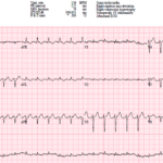
UMMC Cases Part IV: Two Easily Missed ECG Diagnoses
A 73-year-old woman presents to the ED with palpitations and shortness of breath. Her ECG shows a regular narrow-complex tachycardia, and the computer interpretation is accepted by both the emergency…
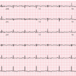
UMMC Cases Part III: When T Waves Help Tell the Story
A 23-year-old woman presents with palpitations and lightheadedness after a spontaneous episode of regular wide complex tachycardia. Her post-conversion ECG shows anterior T wave inversions with a subtle abnormality hiding…
QRS Fragmentation (fQRS)
Key Points: Definition: Fragmented QRS (fQRS) refers to additional notches or deflections within the QRS complex, reflecting heterogeneous ventricular depolarization. In a narrow QRS, classic criteria include an additional R…
Epsilon Waves
Key Points: Definition: Epsilon waves are reproducible, low-amplitude signals occurring between the end of the QRS complex and onset of the T wave, most often seen in the right precordial…
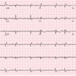
UMMC Cases Part II: Grouped Beats, Slow VT, and Diffuse ST Elevation
A 63-year-old man presents with shortness of breath. His ECG shows an unusual rhythm with repetitive groups of two beats separated by pauses.
T Wave Inversions in V1-V3: DDx
Key Points: Anterior T wave inversion in V1-V3 is not synonymous with “anteroseptal ischemia.” The differential includes ACS, right heart strain, conduction/structural disease, and normal variants. In acute care, the…
Escape-Capture Bigeminy
Key Points: Definition: Escape-capture bigeminy is an uncommon rhythm in which escape beats alternate with conducted sinus capture beats, producing a repeating paired pattern. Mechanism: Sinus slowing, sinus pauses, or…
Inverted P Waves in V1: DDx
Key Points: Know What Is Normal: The sinus P wave in V1 is often biphasic, with an initial positive and terminal negative component. A predominantly or completely negative P wave…
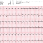
ECG Hallucinations: The Rhythms You See and the Rhythms You Miss
A patient presents with a regular narrow-complex tachycardia. The computerized interpretation identifies sinus tachycardia, and the rhythm appears superficially compatible with that diagnosis when lead II is reviewed. However, closer…
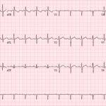
ECG Hallucinations: When S1Q3T3 Misleads
A 51-year-old truck driver presents to the ED after a brief syncopal episode at a rest stop. It is the middle of summer, his truck’s air conditioning is not working,…
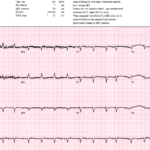
The Rhythm Behind the Irregularity
A 69-year-old woman presenting with sepsis gets the following ECG for tachycardia while febrile and shivering. The baseline is poor, atrial activity is difficult to identify, and the computer interpretation…
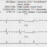
A Closer Look at the J Point
A 72-year-old man undergoes a prehospital 12-lead ECG. The tracing appears to show ST segment elevation in leads III, aVF, and aVR, raising concern for an inferior STEMI or high-risk…
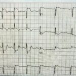
Three High Risk ECGs and the Right Next Move
A 34 year old man presents with acute chest pain radiating to the left arm associated with diaphoresis. He has hyperlipidemia and a family history of early coronary disease. The…
Understanding ECG Artifacts
Key Points: Definition: ECG artifact is a noncardiac signal that distorts, obscures, or mimics the heart’s true electrical activity. Common sources: Patient movement or muscle activity, poor electrode contact, lead…
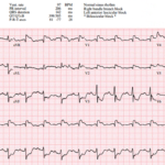
Post-Arrest STEMI: Cath Lab or Caution?
A 72-year-old man is brought to the ED after witnessed cardiac arrest. Bystander CPR was started quickly, EMS found a nonshockable rhythm, epinephrine was given, and ROSC was achieved. Forty…
J Point: Basics
Key Points: The J point is the junction between the end of the QRS complex and the beginning of the ST segment. It is a location, not a separate waveform….
Post-Cardiac Arrest ECGs Hub
Key Points: The first post-ROSC ECG is essential but imperfect. Obtain it immediately, but interpret it in context. Global ischemia, defibrillation, acidosis, hypothermia, vasopressors, artifact, and severe metabolic derangements can…
Post-Arrest STEMI: Cath Lab Decisions
Key Points: Persistent ST elevation after ROSC remains a guideline-supported indication for emergency coronary angiography. The 2025 ACC/AHA/ACEP/NAEMSP/SCAI ACS guideline recommends emergency angiography for patients after cardiac arrest with suspected…
ECG Tags
- 5th Universal Definition of MI
- A-Z
- ACS Mimics
- ACS-OMI
- Activation
- Acute MI
- Advanced Level Curriculum
- Annual ECG Competition
- Anterior OMI
- Anterior STEMI
- Approach
- Arrest
- Arrhythmia
- Arrhythmogenic Cardiomyopathy
- Artifact
- ARVC
- ARVD
- Atrial Parasystole
- Attending
- AV Block
- aVF
- aVL
- aVR
- Axis
- Barcelona
- Basics
- Bix Rule
- Board Review
- Bradyarrhythmia
- Cath Lab
- Chest Pain
- Clumped Beats
- Competition
- Conduction
- Core
- Core Level Curriculum
- Critical ECG Patterns
- Curriculum
- DDx
- Delta
- Devices
- Diagonal Branch Occlusion
- Differential Diagnoses
- Diffuse ST Elevation
- Documentation
- Early Repolarization
- ECG Interpretation
- ECG Localization
- ECG Variant
- Education/Teaching
- Electrolytes
- Emergencies
- Emergent Cath Lab Activation
- EMS
- Expert Level Curriculum
- Flutter
- Flutter Waves
- Foundations Level Curriculum
- Guidelines
- High-Lateral STEMI
- Hub
- Hyperacute T waves
- Hypercalcemia
- Hyperkalemia
- Hypermagnesemia
- Hypocalcemia
- Hypokalemia
- Hypomagnesemia
- Hyponatremia
- Hypothermia
- I
- II
- Index
- Inferior OMI
- Inferior STEMI
- Intervals
- Irregular
- Ischemia
- Ischemia & Infarction
- J point
- J Waves
- JT
- Junctional Rhythm
- Juvenile T wave
- LAD Occlusion
- Lateral STEMI
- LBBB
- LCx Occlusion
- Lead Placement
- Left Atrial Enlargement (LAE)
- Life Savers
- LV an
- LV Aneurysm
- LVAD
- Mastery Level Curriculum
- Metabolic
- MI
- Mimics
- Morphology
- Narrow QRS
- Occlusion MI
- OMI Pattern
- Orthodromic AVRT
- Osborn Waves
- P Wave
- Paced Rhythms
- Pacemaker/ICD
- Paramedics
- Pauses
- PE
- Pediatrics
- Pericardial
- Pericarditis
- PGY-1
- PGY-2
- PGY-3
- PGY-4
- Post-Arrest
- post-arrest STEMI
- Post-Cardiac Arrest
- Posterior Extension
- Posterior MI
- Posterior STEMI
- potpourri
- PR
- Preexcitation
- Premature Complexes
- Prolonged QT
- Pulmonary Embolism
- Pulse-Tapping Artifact
- PVCs
- Q Wave
- QRS
- QT
- R Waves
- RAD
- Rate
- RBBB
- RCA Occlusion
- Regular
- Reperfusion
- Rhythm
- Right Atrial Enlargement (RAE)
- RR
- S Waves
- S1Q3T3
- Segments
- Seizure
- Serial ECGs
- Sgarbossa
- Shock
- South African Flag Sign
- ST
- ST Depression
- ST Elevation
- STEMI
- STEMI Equivalent
- STEMI Mimics
- STEMI Negative OMI
- Stepwise
- Stroke
- Structural
- Students
- SVT
- Syncope
- T Wave Inversion
- T Waves
- Tachyarrhythmia
- Tachycardia
- Tamponade
- Terminal QRS Distortion
- Toxicology
- TP
- Traditional STEMI Criteria
- U Waves
- Unstable
- V1-V4
- V2
- V5
- V6
- Vectors
- Ventricular Repolarization
- Ventricular Rhythms
- Ventricular Tachycardia
- Voltage
- Waveforms
- Wide QRS
- Workflow
- WPW
© 2026 ECG Weekly. All rights reserved. | Terms of Use | Privacy Policy
Powered by the member(dev) platform
 Loading...