
ECG Interpretation
Results:

The Life Savers: Critical ECGs Hub
Key Points: Life Savers are the can’t-miss ECGs. These patterns may reflect immediately life-threatening ischemic, electrical, mechanical, obstructive, toxicologic, or metabolic emergencies. This hub is built for rapid action. Use…

Stepwise Approach to STAT ECGs Hub
Key Points: Use a consistent ECG routine. In acute care, a repeatable approach reduces misses and helps you recognize dangerous patterns faster. Prioritize life threats first. Instability, malignant rhythms, occlusion…
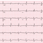
The Syncope ECG With Too Much P
A 68-year-old man has syncope, then has a second syncopal episode while lying still on a stretcher during evaluation at an outpatient clinic. He is sent emergently to the ED….

Preexcitation Pitfalls (Part 4): Potpourri Cases & Final Teaching Points
A 49-year-old man arrives with palpitations and chest discomfort. The monitor shows an irregular, wide-complex tachycardia with varying morphology and rates nearing 250 to 300 bpm. The team debates polymorphic…
QRS Morphology and ST-T Interpretation: Basics
Key Points: Read the QRS before you read the ST segment or T wave. Ventricular depolarization shapes repolarization. Narrow QRS usually reflects normal His-Purkinje conduction. Wide QRS suggests abnormal ventricular…
Preexcitation Syndromes: Overview
Key Points: Pre-excitation means an accessory pathway allows atrial impulses to reach the ventricle without traversing the AV node, producing early ventricular activation. A delta wave is the defining ECG…
Short QT Interval: DDx
Key Points: Short QT Interval: A QT interval is considered short when the corrected QT (QTc) interval is less than 350 ms. A short QT interval on the ECG can…
Short QT Syndrome
Key Points: Short QT Syndrome (SQTS) is a rare condition characterized by a shortened QT interval on the ECG, increasing the risk of atrial and ventricular arrhythmias, including sudden cardiac…
P Wave: Basics
Key Points: Definition and measurement: The P wave is atrial depolarization. Measure duration from initial deflection to return to baseline and amplitude from baseline to peak. Normal values: Duration <120…
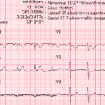
Baltimore City EMS ECGs: Pitfalls and Mimics (Part 1)
A 68-year-old man is brought to the emergency department by EMS with acute chest discomfort. The following prehospital ECG was obtained and shows concave ST elevation across multiple leads. The…
Waveforms, Segments, & Intervals: Basics
Key Points: Every ECG tracing is built from waveforms (deflections), segments (baseline portions between waveforms), and intervals (time that include waveforms plus segments). Waveforms describe electrical events (depolarization or repolarization)….
ECG STAT Curriculum Hub
Why ECG Interpretation Matters: The ECG is one of the fastest, highest-impact diagnostic tests in acute care. When interpretation is rapid and accurate, patients get timely reperfusion, pacing, defibrillation, antidotes,…
ECG Findings of LV Aneurysm
Key Points: Definition: A true LV aneurysm is a chronic, post transmural MI complication from scarred myocardium with akinetic or dyskinetic (paradoxical) wall motion. ECG hallmark: Persistent ST elevation in…
Post-Thrombolytic Reperfusion ECG Findings
Key Points Reperfusion after fibrinolysis is a bedside diagnosis using a bundle of findings: symptoms, ECG trend, and hemodynamic/electrical stability. Best ECG marker of successful fibrinolysis: at least 50% ST-segment…
ECG Evidence of Reperfusion After Occlusion
Key Points: Reperfusion and re-occlusion can occur spontaneously or after therapy. The ECG often reflects these changes earlier than symptoms. Most useful bedside ECG marker of reperfusion is ST-segment resolution…
Pulse-Tapping Artifact
Key Points: Mechanical artifact caused by an ECG electrode sitting on top of a strong arterial pulse. Seen frequently in dialysis patients with AV fistulas. Can mimic serious pathology including…
Premature Junctional Complexes (PJCs)
Key Points PJCs are premature impulses from ectopic foci in or near the AV junction. ECG hallmark is a narrow premature beat with an absent or retrograde P wave. Retrograde…
J Wave: Basics
Key Points Definitions: The J point is the QRS–ST junction (a location). A J wave is an added deflection at or just after that point (notch or slur). Why it…
Proper ECG Electrode Placement
Key Points A diagnostic 12‑lead ECG is only as good as your electrode placement. Misplacement can mimic MI, BBB, or poor R‑wave progression and trigger unnecessary workups. A standard 12‑lead…
ECG Findings in Tension Pneumothorax
Key Points Tension pneumothorax is a clinical diagnosis—ECG changes are nonspecific but can provide supportive evidence in the right context. Electrical changes are primarily due to mechanical effects of air…
ECG Tags
- 5th Universal Definition of MI
- A-Z
- ACS Mimics
- ACS-OMI
- Activation
- Acute MI
- Advanced Level Curriculum
- Annual ECG Competition
- Anterior OMI
- Anterior STEMI
- Approach
- Arrest
- Arrhythmia
- Arrhythmogenic Cardiomyopathy
- Artifact
- ARVC
- ARVD
- Atrial Parasystole
- Attending
- AV Block
- aVF
- aVL
- aVR
- Axis
- Barcelona
- Basics
- Bix Rule
- Board Review
- Bradyarrhythmia
- Cath Lab
- Chest Pain
- Clumped Beats
- Competition
- Conduction
- Core
- Core Level Curriculum
- Critical ECG Patterns
- Curriculum
- DDx
- Delta
- Devices
- Diagonal Branch Occlusion
- Differential Diagnoses
- Diffuse ST Elevation
- Documentation
- Early Repolarization
- ECG Interpretation
- ECG Localization
- ECG Variant
- Education/Teaching
- Electrolytes
- Emergencies
- Emergent Cath Lab Activation
- EMS
- Expert Level Curriculum
- Flutter
- Flutter Waves
- Foundations Level Curriculum
- Guidelines
- High-Lateral STEMI
- Hub
- Hyperacute T waves
- Hypercalcemia
- Hyperkalemia
- Hypermagnesemia
- Hypocalcemia
- Hypokalemia
- Hypomagnesemia
- Hyponatremia
- Hypothermia
- I
- II
- Index
- Inferior OMI
- Inferior STEMI
- Intervals
- Irregular
- Ischemia
- Ischemia & Infarction
- J point
- J Waves
- JT
- Junctional Rhythm
- Juvenile T wave
- LAD Occlusion
- Lateral STEMI
- LBBB
- LCx Occlusion
- Lead Placement
- Left Atrial Enlargement (LAE)
- Life Savers
- LV an
- LV Aneurysm
- LVAD
- Mastery Level Curriculum
- Metabolic
- MI
- Mimics
- Morphology
- Narrow QRS
- Occlusion MI
- OMI Pattern
- Orthodromic AVRT
- Osborn Waves
- P Wave
- Paced Rhythms
- Pacemaker/ICD
- Paramedics
- Pauses
- PE
- Pediatrics
- Pericardial
- Pericarditis
- PGY-1
- PGY-2
- PGY-3
- PGY-4
- Post-Arrest
- post-arrest STEMI
- Post-Cardiac Arrest
- Posterior Extension
- Posterior MI
- Posterior STEMI
- potpourri
- PR
- Preexcitation
- Premature Complexes
- Prolonged QT
- Pulmonary Embolism
- Pulse-Tapping Artifact
- PVCs
- Q Wave
- QRS
- QT
- R Waves
- RAD
- Rate
- RBBB
- RCA Occlusion
- Regular
- Reperfusion
- Rhythm
- Right Atrial Enlargement (RAE)
- RR
- S Waves
- S1Q3T3
- Segments
- Seizure
- Serial ECGs
- Sgarbossa
- Shock
- South African Flag Sign
- ST
- ST Depression
- ST Elevation
- STEMI
- STEMI Equivalent
- STEMI Mimics
- STEMI Negative OMI
- Stepwise
- Stroke
- Structural
- Students
- SVT
- Syncope
- T Wave Inversion
- T Waves
- Tachyarrhythmia
- Tachycardia
- Tamponade
- Terminal QRS Distortion
- Toxicology
- TP
- Traditional STEMI Criteria
- U Waves
- Unstable
- V1-V4
- V2
- V5
- V6
- Vectors
- Ventricular Repolarization
- Ventricular Rhythms
- Ventricular Tachycardia
- Voltage
- Waveforms
- Wide QRS
- Workflow
- WPW
© 2026 ECG Weekly. All rights reserved. | Terms of Use | Privacy Policy
Powered by the member(dev) platform
 Loading...