Emergencies
Results:
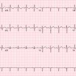
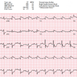
ECG Hallucinations: When S1Q3T3 Misleads
A 51-year-old truck driver presents to the ED after a brief syncopal episode at a rest stop. It is the middle of summer, his truck’s air conditioning is not working,…

S1Q3T3 Pattern
Key Points: Not a PE diagnosis: S1Q3T3 is classically associated with pulmonary embolism, but it is neither sensitive nor specific enough to diagnose or exclude PE. Think acute right heart…
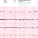
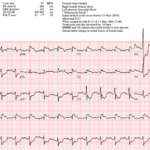
ECG in Pulmonary Embolism (PE)
Key Points: The ECG cannot rule in or rule out PE: No single ECG finding is sufficiently sensitive or specific to diagnose or exclude acute pulmonary embolism. A Normal ECG…
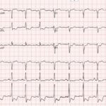
High Risk Pulmonary Embolism (PE)
Key Points: The ECG can provide an early warning of severe RV strain: No ECG finding defines high risk PE, but marked or evolving signs of acute RV pressure overload…
The Rhythm Behind the Irregularity
A 69-year-old woman presenting with sepsis gets the following ECG for tachycardia while febrile and shivering. The baseline is poor, atrial activity is difficult to identify, and the computer interpretation…
Four ECGs Hiding a High-Risk Coronary Pattern
A 56-year-old man presents with 1 week of concerning chest pain and shortness of breath. His ECG does not meet conventional STEMI criteria, but it shows subtle ST-segment abnormalities in…
Three High Risk ECGs and the Right Next Move
A 34 year old man presents with acute chest pain radiating to the left arm associated with diaphoresis. He has hyperlipidemia and a family history of early coronary disease. The…
Barcelona Criteria in Left Bundle Branch Block (LBBB): OMI Pattern
Key Points: The Barcelona Criteria are ECG criteria proposed to identify acute myocardial infarction in patients with LBBB. Core problem: LBBB causes expected secondary ST-T changes, so standard STEMI millimeter…
Aslanger Pattern: OMI Pattern
Key Points: Aslanger pattern is an OMI pattern that can identify acute inferior occlusion despite not meeting traditional STEMI criteria. The key finding is ST segment elevation isolated to lead…
Occlusion MI in Left Bundle Branch Block (LBBB): STEMI Equivalent Pattern
Key Points: LBBB does not exclude acute coronary occlusion. LBBBs produce abnormal depolarization and expected secondary ST-T changes, which can mask or mimic ischemia. Acute occlusion MI can still be…
Post-Arrest STEMI: Cath Lab or Caution?
A 72-year-old man is brought to the ED after witnessed cardiac arrest. Bystander CPR was started quickly, EMS found a nonshockable rhythm, epinephrine was given, and ROSC was achieved. Forty…
Post-Cardiac Arrest ECGs Hub
Key Points: The first post-ROSC ECG is essential but imperfect. Obtain it immediately, but interpret it in context. Global ischemia, defibrillation, acidosis, hypothermia, vasopressors, artifact, and severe metabolic derangements can…
Post-Arrest STEMI: Cath Lab Decisions
Key Points: Persistent ST elevation after ROSC remains a guideline-supported indication for emergency coronary angiography. The 2025 ACC/AHA/ACEP/NAEMSP/SCAI ACS guideline recommends emergency angiography for patients after cardiac arrest with suspected…
Post-Arrest No STEMI: When Cath Can Wait
Key Points: Stable post-arrest patients without ST elevation should not go to reflex immediate cath solely because cardiac arrest occurred. Randomized trials in OHCA patients without ST elevation have not…
Pediatric ECG in Cardiac Arrest
Key Points: Pediatric arrest is usually respiratory, hypoxic, or shock-related, not primary coronary occlusion. The ECG still matters because it can reveal reversible metabolic, toxicologic, structural, inflammatory, or inherited electrical…
A Post-Arrest ECG With a Dangerous New Bundle Branch Block
A 72-year-old man is brought to the ED after a witnessed out-of-hospital cardiac arrest. Bystander CPR is started quickly, EMS finds a non-shockable rhythm, and ROSC is achieved after one…
The Cath Lab Was Activated, But Something Didn’t Fit
A 70-year-old woman with CHF, COPD, intermittent atrial fibrillation, chronic pain medication use, and recent poor intake develops sudden dyspnea at rest and is found somnolent and bradycardic in the…
Sinus Node Dysfunction (Sick Sinus Syndrome & Bradycardia-Tachycardia Syndrome)
Key Points: A spectrum, not a single rhythm: Sinus node dysfunction includes inappropriate sinus bradycardia, sinus pauses or arrest, SA exit block, chronotropic incompetence, and alternating atrial tachyarrhythmias with bradycardia….
Sinoatrial Exit Block
Key Points: Definition: Sinoatrial, or SA, exit block occurs when the sinus node generates an impulse that fails to conduct into the surrounding atrial tissue. The blocked impulse produces no…
Sinus Arrest
Key Points: Definition: Sinus arrest occurs when the sinus node temporarily fails to generate an impulse. This produces an absence of the expected P wave and its associated QRS complex….
ECG Tags
- A-Z
- ACS Mimics
- ACS-OMI
- Activation
- Advanced Level Curriculum
- Annual ECG Competition
- Anterior OMI
- Anterior STEMI
- Approach
- Arrest
- Arrhythmia
- Arrhythmogenic Cardiomyopathy
- Artifact
- ARVC
- ARVD
- Atrial Parasystole
- Attending
- AV Block
- aVF
- aVL
- aVR
- Axis
- Barcelona
- Basics
- Bix Rule
- Board Review
- Bradyarrhythmia
- Cath Lab
- Chest Pain
- Clumped Beats
- Competition
- Conduction
- Core
- Core Level Curriculum
- Critical ECG Patterns
- Curriculum
- DDx
- Delta
- Devices
- Diagonal Branch Occlusion
- Differential Diagnoses
- Diffuse ST Elevation
- Documentation
- Early Repolarization
- ECG Interpretation
- ECG Localization
- ECG Variant
- Education/Teaching
- Electrolytes
- Emergencies
- Emergent Cath Lab Activation
- EMS
- Expert Level Curriculum
- Flutter
- Flutter Waves
- Foundations Level Curriculum
- Guidelines
- High-Lateral STEMI
- Hub
- Hyperacute T waves
- Hypercalcemia
- Hyperkalemia
- Hypermagnesemia
- Hypocalcemia
- Hypokalemia
- Hypomagnesemia
- Hyponatremia
- Hypothermia
- I
- II
- Index
- Inferior OMI
- Inferior STEMI
- Intervals
- Irregular
- Ischemia
- Ischemia & Infarction
- J point
- J Waves
- JT
- Junctional Rhythm
- Juvenile T wave
- LAD Occlusion
- Lateral STEMI
- LBBB
- LCx Occlusion
- Lead Placement
- Left Atrial Enlargement (LAE)
- Life Savers
- LV an
- LV Aneurysm
- LVAD
- Mastery Level Curriculum
- Metabolic
- Mimics
- Morphology
- Narrow QRS
- Occlusion MI
- OMI Pattern
- Orthodromic AVRT
- Osborn Waves
- P Wave
- Paced Rhythms
- Pacemaker/ICD
- Paramedics
- Pauses
- PE
- Pediatrics
- Pericardial
- Pericarditis
- PGY-1
- PGY-2
- PGY-3
- PGY-4
- Post-Arrest
- post-arrest STEMI
- Post-Cardiac Arrest
- Posterior Extension
- Posterior MI
- Posterior STEMI
- potpourri
- PR
- Preexcitation
- Premature Complexes
- Prolonged QT
- Pulmonary Embolism
- Pulse-Tapping Artifact
- PVCs
- Q Wave
- QRS
- QT
- R Waves
- RAD
- Rate
- RBBB
- RCA Occlusion
- Regular
- Reperfusion
- Rhythm
- Right Atrial Enlargement (RAE)
- RR
- S Waves
- S1Q3T3
- Segments
- Seizure
- Serial ECGs
- Sgarbossa
- Shock
- South African Flag Sign
- ST
- ST Depression
- ST Elevation
- STEMI
- STEMI Equivalent
- STEMI Mimics
- STEMI Negative OMI
- Stepwise
- Stroke
- Structural
- Students
- SVT
- Syncope
- T Wave Inversion
- T Waves
- Tachyarrhythmia
- Tachycardia
- Tamponade
- Terminal QRS Distortion
- Toxicology
- TP
- Traditional STEMI Criteria
- U Waves
- Unstable
- V1-V4
- V2
- V5
- V6
- Vectors
- Ventricular Repolarization
- Ventricular Rhythms
- Ventricular Tachycardia
- Voltage
- Waveforms
- Wide QRS
- Workflow
- WPW
© 2026 ECG Weekly. All rights reserved. | Terms of Use | Privacy Policy
Powered by the member(dev) platform
 Loading...