Emergencies
Results:
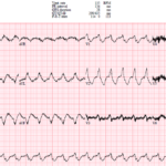
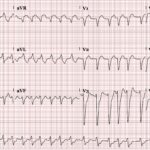
UMEM Cases, Part 4: When the Computer Misses the Rhythm and Flutter Fakes a STEMI
A 44-year-old man with severe cardiomyopathy, an LVAD, chronic amiodarone therapy, and an AICD presents with palpitations. His ECG shows a regular wide-complex tachycardia, but the rate is only 135….

New RBBB + LAFB (Bifascicular Block) in ACS: OMI Pattern
Key Points: In a patient with ischemic symptoms, new RBBB + LAFB should raise concern for proximal LAD or septal ischemia until proven otherwise, especially if the patient has ongoing…
Wide QRS Complex: DDx
Key Points: A QRS duration greater than 120 ms reflects delayed or abnormal ventricular depolarization. A wide QRS may be chronic and benign in context, or it may be the…
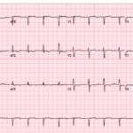
UMEM Cases, Part 3: When the Diagnosis Seems Clear and When It Is Not
A 71-year-old man presents with shortness of breath, and his ECG is initially read as a junctional rhythm. On later review, it is even mistaken for atrial fibrillation. But the…
Dysrhythmias in LVAD Patients
Key Points: Continuous-flow LVADs can mask cardiovascular collapse. Patients may remain awake during sustained VT or even VF because the pump can provide temporary flow. Treat the rhythm and the…
Pediatric ECG Red Flags
Key Points: Any wide QRS (>90 ms) in an infant or small child is abnormal and should trigger evaluation for VT, sodium-channel blockade, or conduction disease. QTc >450 ms in…
Unstable Bradyarrhythmias
Key Points: Unstable bradyarrhythmias cause poor perfusion which can rapidly progress to shock, irreversible organ injury, or cardiac arrest. Priority: Do not treat the heart rate alone. Treat clinical instability….
Severe Hypothermia
Key Points: Severe hypothermia causes predictable ECG slowing and conduction delay. Sinus bradycardia, PR/QRS/QT prolongation, and atrial fibrillation with a slow ventricular response are common as core temperature falls. Osborn…
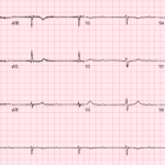
UMEM Cases, Part 2: When the ECG Conceals and When It Reveals
An 81-year-old woman presents with lightheadedness and marked bradycardia. Her ECG shows more P waves than QRS complexes, but the mechanism is not immediately clear. The key question is whether…
Computer Interpreted “Normal” ECGs
Key Points: Do not trust a computer read of “normal” without your own review. Computer interpretation is especially unreliable for subtle or early ischemia, including hyperacute T waves, minimal ST…
Torsade de Pointes (TdP)
Key Points: Definition: Torsade de pointes is a specific subtype of polymorphic ventricular tachycardia that occurs in the setting of QT prolongation. ECG pattern: TdP shows beat-to-beat variation in QRS…
ST Elevation in aVR with Diffuse ST Segment Depression: OMI Pattern
Key Points: Pattern, not a STEMI equivalent. ST elevation in aVR (≥1 mm), often with ST elevation in V1 and diffuse ST depression (≥1 mm in ≥6 leads), represents high-risk…
Hyperkalemia Emergencies
Key Points: Severe Hyperkalemia Mimics Several Life-Threatening Conditions: Severe hyperkalemia is one of the most dangerous ECG mimics in emergency medicine. It can resemble unstable bradyarrhythmias, VT, STEMI, and pacemaker…
Polymorphic Ventricular Tachycardia (PMVT)
Key Points: Definition: Polymorphic ventricular tachycardia is VT with beat-to-beat variation in QRS morphology, axis, and amplitude. Clinical significance: PMVT is electrically unstable and can rapidly deteriorate into ventricular fibrillation…
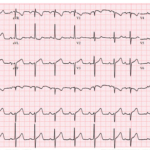
UMEM Cases, Part 1: When the ECG Lies and When It Evolves
A 72-year-old man presents with chest pain and shortness of breath. His ECG shows sinus rhythm with LVH, mild inferior ST elevation, and lateral ST-T abnormalities that some interpret as…
Nonsustained Ventricular Tachycardia (NSVT)
Key Points: Definition: Nonsustained ventricular tachycardia is 3 or more consecutive ventricular beats lasting less than 30 seconds and terminating spontaneously. Rate: VT is usually faster than 120 bpm, but…
STEMI vs Wide QRS (BBBs & Paced Rhythms): DDx
Key Points: Wide QRS rhythms distort repolarization. Bundle branch block and ventricular pacing create secondary ST-T changes even without occlusion MI. Appropriate discordance is expected. ST segments and T waves…
South African Flag Sign: OMI Pattern
Key Points: Pattern: The South African Flag sign is the combination of ST elevation in I, aVL, and V2 with reciprocal ST depression in III. It is a subtle but…
Three ECG Traps You Cannot Afford to Miss
A 60-year-old woman presents with palpitations and an irregular wide-complex tachycardia. The computer calls atrial fibrillation with a left bundle branch block, but a subtle clue in the precordial leads…
Occlusion MI in Ventricular Paced Rhythms: STEMI Equivalent Pattern
Key Points: Ventricular paced rhythms can mask acute coronary occlusion. Pacing alters depolarization and produces expected secondary ST-T abnormalities, so standard STEMI criteria are unreliable. Appropriate discordance is expected in…
ECG Tags
- A-Z
- ACS Mimics
- ACS-OMI
- Activation
- Advanced Level Curriculum
- Annual ECG Competition
- Anterior OMI
- Anterior STEMI
- Approach
- Arrest
- Arrhythmia
- Arrhythmogenic Cardiomyopathy
- Artifact
- ARVC
- ARVD
- Atrial Parasystole
- Attending
- AV Block
- aVF
- aVL
- aVR
- Axis
- Barcelona
- Basics
- Bix Rule
- Board Review
- Bradyarrhythmia
- Cath Lab
- Chest Pain
- Clumped Beats
- Competition
- Conduction
- Core
- Core Level Curriculum
- Critical ECG Patterns
- Curriculum
- DDx
- Delta
- Devices
- Diagonal Branch Occlusion
- Differential Diagnoses
- Diffuse ST Elevation
- Documentation
- Early Repolarization
- ECG Interpretation
- ECG Localization
- ECG Variant
- Education/Teaching
- Electrolytes
- Emergencies
- Emergent Cath Lab Activation
- EMS
- Expert Level Curriculum
- Flutter
- Flutter Waves
- Foundations Level Curriculum
- Guidelines
- High-Lateral STEMI
- Hub
- Hyperacute T waves
- Hypercalcemia
- Hyperkalemia
- Hypermagnesemia
- Hypocalcemia
- Hypokalemia
- Hypomagnesemia
- Hyponatremia
- Hypothermia
- I
- II
- Index
- Inferior OMI
- Inferior STEMI
- Intervals
- Irregular
- Ischemia
- Ischemia & Infarction
- J point
- J Waves
- JT
- Junctional Rhythm
- Juvenile T wave
- LAD Occlusion
- Lateral STEMI
- LBBB
- LCx Occlusion
- Lead Placement
- Left Atrial Enlargement (LAE)
- Life Savers
- LV an
- LV Aneurysm
- LVAD
- Mastery Level Curriculum
- Metabolic
- Mimics
- Morphology
- Narrow QRS
- Occlusion MI
- OMI Pattern
- Orthodromic AVRT
- Osborn Waves
- P Wave
- Paced Rhythms
- Pacemaker/ICD
- Paramedics
- Pauses
- PE
- Pediatrics
- Pericardial
- Pericarditis
- PGY-1
- PGY-2
- PGY-3
- PGY-4
- Post-Arrest
- post-arrest STEMI
- Post-Cardiac Arrest
- Posterior Extension
- Posterior MI
- Posterior STEMI
- potpourri
- PR
- Preexcitation
- Premature Complexes
- Prolonged QT
- Pulmonary Embolism
- Pulse-Tapping Artifact
- PVCs
- Q Wave
- QRS
- QT
- R Waves
- RAD
- Rate
- RBBB
- RCA Occlusion
- Regular
- Reperfusion
- Rhythm
- Right Atrial Enlargement (RAE)
- RR
- S Waves
- S1Q3T3
- Segments
- Seizure
- Serial ECGs
- Sgarbossa
- Shock
- South African Flag Sign
- ST
- ST Depression
- ST Elevation
- STEMI
- STEMI Equivalent
- STEMI Mimics
- STEMI Negative OMI
- Stepwise
- Stroke
- Structural
- Students
- SVT
- Syncope
- T Wave Inversion
- T Waves
- Tachyarrhythmia
- Tachycardia
- Tamponade
- Terminal QRS Distortion
- Toxicology
- TP
- Traditional STEMI Criteria
- U Waves
- Unstable
- V1-V4
- V2
- V5
- V6
- Vectors
- Ventricular Repolarization
- Ventricular Rhythms
- Ventricular Tachycardia
- Voltage
- Waveforms
- Wide QRS
- Workflow
- WPW
© 2026 ECG Weekly. All rights reserved. | Terms of Use | Privacy Policy
Powered by the member(dev) platform
 Loading...