
Emergencies
Results:

Atrial Flutter 2:1 Conduction
Key Points: Atrial flutter is a macro-reentrant atrial tachycardia, most commonly typical cavotricuspid isthmus-dependent right atrial flutter, with an atrial rate usually near 300 bpm. With 2:1 AV conduction, the…
Atrial Flutter 1:1 Conduction
Key Points: Rare, high-risk rhythm. Atrial flutter with 1:1 conduction can produce ventricular rates of 240-320 bpm and may rapidly cause hypotension, ischemia, or collapse. Often mimics VT. The QRS…
Advanced (High-Grade) AV Block
Key Points: Advanced or high-grade AV block is a severe second-degree AV block with 2 or more consecutive non-conducted P waves, such as 3:1 or 4:1 conduction. Do not force…
Preexcitation Syndromes: Overview
Key Points: Pre-excitation means an accessory pathway allows atrial impulses to reach the ventricle without traversing the AV node, producing early ventricular activation. A delta wave is the defining ECG…
Wolff-Parkinson White (WPW) Syndrome
Key Points: Pattern vs syndrome: WPW pattern is ECG evidence of pre-excitation without symptoms. WPW syndrome is pattern plus symptomatic tachyarrhythmia (palpitations, syncope, “seizure”, aborted sudden cardiac arrest). PR interval…
Short QT Syndrome
Key Points: Short QT Syndrome (SQTS) is a rare condition characterized by a shortened QT interval on the ECG, increasing the risk of atrial and ventricular arrhythmias, including sudden cardiac…
WPW Syndrome and Pseudo-MI Patterns
Key Points: WPW alters ventricular depolarization, producing secondary repolarization abnormalities that can mimic or mask myocardial infarction. ST-segment deviation in WPW is often non-ischemic, driven by abnormal activation via the…
Atrial Fibrillation with WPW (Pre-excited AF)
Key Points: Pre-excited AF is the most dangerous WPW rhythm. It can deteriorate quickly to VF because the accessory pathway may conduct atrial impulses to the ventricle at extreme rates….
WPW with Antidromic SVT (Antidromic AVRT)
Key Points: Antidromic AVRT is an AV re-entrant tachycardia that conducts antegrade down the accessory pathway and returns retrograde through the AV node (or another pathway), producing a regular wide-complex…
WPW with Orthodromic SVT (Orthodromic AVRT)
Key Points: Orthodromic AVRT is the most common tachyarrhythmia in WPW and presents as a regular narrow-complex SVT that is indistinguishable from AVNRT during the tachycardia. Mechanism: antegrade conduction down…
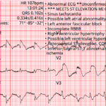
Baltimore City EMS ECGs: Pitfalls and Mimics (Part 2)
A 54-year-old man presents to the emergency department by EMS with acute shortness of breath. A prehospital ECG triggers a STEMI alert based on the computer interpretation. The tracing shows…
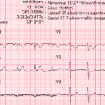
Baltimore City EMS ECGs: Pitfalls and Mimics (Part 1)
A 68-year-old man is brought to the emergency department by EMS with acute chest discomfort. The following prehospital ECG was obtained and shows concave ST elevation across multiple leads. The…
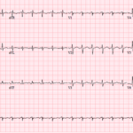
Three More ECG Pitfalls That Punish Anchoring Bias
A 51-year-old man with lung cancer presents with shortness of breath and tachycardia. The arrival ECG shows an S1Q3 pattern and seems to support a familiar diagnosis that would normally…
Bidirectional Ventricular Tachycardia (BiVT)
Key Points: BiVT is a regular wide-complex tachycardia with strict beat-to-beat alternation of QRS axis and/or bundle-branch pattern (often an approximately 180° frontal-plane axis flip). In adults, assume digoxin toxicity…
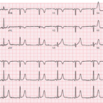
Four ECG Pitfalls That Punish Anchoring Bias
A 43-year-old woman with sharp left-sided chest pain and minimal cardiac risk factors has an initial ECG that is not diagnostic for STEMI. She looks stable, but one feature on…
STEMI (-) Occlusion MI: OMI Patterns
Key Points: Traditional STEMI criteria miss many acute coronary occlusions. A substantial proportion of true OMIs do not meet classic millimetric STEMI thresholds. OMI is a pathophysiologic diagnosis, not an…
Hypokalemic Periodic Paralysis
Key Points: Definition: Hypokalemic periodic paralysis (HPP) is a rare disorder characterized by episodic, reversible flaccid muscle weakness associated with a shift of potassium from the extracellular space into skeletal…
STEMI in the Presence of Baseline ECG Abnormalities
Key Points: Baseline ECG abnormalities do not protect patients from occlusion MI. They increase miss rates because they distort the ST segment and T waves. The core question is not…
Lateral and High-Lateral STEMI: Criteria, Localization, and Pitfalls
Key Points: Lateral and high-lateral STEMI often present with subtle ST elevation and are commonly missed or labeled as nonspecific ST-T changes. Small-appearing ECG changes may represent true coronary occlusion…
Inferior STEMI: Criteria, RV Involvement, and Pitfalls
Key Points: Inferior STEMI is the most common STEMI subtype and is frequently complicated by right ventricular and posterior involvement. Inferior occlusion may present with classic ST elevation, subtle ischemic…
ECG Tags
- 5th Universal Definition of MI
- A-Z
- ACS Mimics
- ACS-OMI
- Activation
- Acute MI
- Advanced Level Curriculum
- Annual ECG Competition
- Anterior OMI
- Anterior STEMI
- Approach
- Arrest
- Arrhythmia
- Arrhythmogenic Cardiomyopathy
- Artifact
- ARVC
- ARVD
- Atrial Parasystole
- Attending
- AV Block
- aVF
- aVL
- aVR
- Axis
- Barcelona
- Basics
- Bix Rule
- Board Review
- Bradyarrhythmia
- Cath Lab
- Chest Pain
- Clumped Beats
- Competition
- Conduction
- Core
- Core Level Curriculum
- Critical ECG Patterns
- Curriculum
- DDx
- Delta
- Devices
- Diagonal Branch Occlusion
- Differential Diagnoses
- Diffuse ST Elevation
- Documentation
- Early Repolarization
- ECG Interpretation
- ECG Localization
- ECG Variant
- Education/Teaching
- Electrolytes
- Emergencies
- Emergent Cath Lab Activation
- EMS
- Expert Level Curriculum
- Flutter
- Flutter Waves
- Foundations Level Curriculum
- Guidelines
- High-Lateral STEMI
- Hub
- Hyperacute T waves
- Hypercalcemia
- Hyperkalemia
- Hypermagnesemia
- Hypocalcemia
- Hypokalemia
- Hypomagnesemia
- Hyponatremia
- Hypothermia
- I
- II
- Index
- Inferior OMI
- Inferior STEMI
- Intervals
- Irregular
- Ischemia
- Ischemia & Infarction
- J point
- J Waves
- JT
- Junctional Rhythm
- Juvenile T wave
- LAD Occlusion
- Lateral STEMI
- LBBB
- LCx Occlusion
- Lead Placement
- Left Atrial Enlargement (LAE)
- Life Savers
- LV an
- LV Aneurysm
- LVAD
- Mastery Level Curriculum
- Metabolic
- MI
- Mimics
- Morphology
- Narrow QRS
- Occlusion MI
- OMI Pattern
- Orthodromic AVRT
- Osborn Waves
- P Wave
- Paced Rhythms
- Pacemaker/ICD
- Paramedics
- Pauses
- PE
- Pediatrics
- Pericardial
- Pericarditis
- PGY-1
- PGY-2
- PGY-3
- PGY-4
- Post-Arrest
- post-arrest STEMI
- Post-Cardiac Arrest
- Posterior Extension
- Posterior MI
- Posterior STEMI
- potpourri
- PR
- Preexcitation
- Premature Complexes
- Prolonged QT
- Pulmonary Embolism
- Pulse-Tapping Artifact
- PVCs
- Q Wave
- QRS
- QT
- R Waves
- RAD
- Rate
- RBBB
- RCA Occlusion
- Regular
- Reperfusion
- Rhythm
- Right Atrial Enlargement (RAE)
- RR
- S Waves
- S1Q3T3
- Segments
- Seizure
- Serial ECGs
- Sgarbossa
- Shock
- South African Flag Sign
- ST
- ST Depression
- ST Elevation
- STEMI
- STEMI Equivalent
- STEMI Mimics
- STEMI Negative OMI
- Stepwise
- Stroke
- Structural
- Students
- SVT
- Syncope
- T Wave Inversion
- T Waves
- Tachyarrhythmia
- Tachycardia
- Tamponade
- Terminal QRS Distortion
- Toxicology
- TP
- Traditional STEMI Criteria
- U Waves
- Unstable
- V1-V4
- V2
- V5
- V6
- Vectors
- Ventricular Repolarization
- Ventricular Rhythms
- Ventricular Tachycardia
- Voltage
- Waveforms
- Wide QRS
- Workflow
- WPW
© 2026 ECG Weekly. All rights reserved. | Terms of Use | Privacy Policy
Powered by the member(dev) platform
 Loading...