
Tachyarrhythmia
Results:
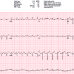
UMMC Cases Part IV: Two Easily Missed ECG Diagnoses
A 73-year-old woman presents to the ED with palpitations and shortness of breath. Her ECG shows a regular narrow-complex tachycardia, and the computer interpretation is accepted by both the emergency…

Monomorphic Ventricular Tachycardia (MMVT)
Key Points Definition: Monomorphic ventricular tachycardia is a ventricular rhythm with a wide QRS and a single, uniform QRS morphology throughout the run. Sustained VT: VT is sustained when it…
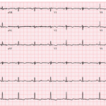
UMMC Cases Part III: When T Waves Help Tell the Story
A 23-year-old woman presents with palpitations and lightheadedness after a spontaneous episode of regular wide complex tachycardia. Her post-conversion ECG shows anterior T wave inversions with a subtle abnormality hiding…
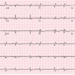
UMMC Cases Part II: Grouped Beats, Slow VT, and Diffuse ST Elevation
A 63-year-old man presents with shortness of breath. His ECG shows an unusual rhythm with repetitive groups of two beats separated by pauses.
Slow Ventricular Tachycardia (VT)
Key Points: Definition: Slow VT refers to ventricular tachycardia occurring at an unusually low ventricular rate, often near or even below the conventional tachycardia threshold. There is no universally accepted…
Ventricular Tachycardia (VT) Mimics
Key Points: Default assumption: A regular wide-complex tachycardia should be considered VT until another diagnosis is more likely. Rate modifies the differential, not the diagnosis. Typical monomorphic VT is often…
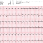
ECG Hallucinations: The Rhythms You See and the Rhythms You Miss
A patient presents with a regular narrow-complex tachycardia. The computerized interpretation identifies sinus tachycardia, and the rhythm appears superficially compatible with that diagnosis when lead II is reviewed. However, closer…
Sinus Node Dysfunction (Sick Sinus Syndrome & Bradycardia-Tachycardia Syndrome)
Key Points: A spectrum, not a single rhythm: Sinus node dysfunction includes inappropriate sinus bradycardia, sinus pauses or arrest, SA exit block, chronotropic incompetence, and alternating atrial tachyarrhythmias with bradycardia….
UMEM Cases, Part 4: When the Computer Misses the Rhythm and Flutter Fakes a STEMI
A 44-year-old man with severe cardiomyopathy, an LVAD, chronic amiodarone therapy, and an AICD presents with palpitations. His ECG shows a regular wide-complex tachycardia, but the rate is only 135….
Junctional Tachycardia
Key Points: Junctional tachycardia is an uncommon supraventricular tachycardia arising from the AV junction, usually due to enhanced automaticity rather than reentry. It is usually a regular narrow-complex tachycardia, although…
Torsade de Pointes (TdP)
Key Points: Definition: Torsade de pointes is a specific form of polymorphic ventricular tachycardia (PMVT) occurring in the setting of prolonged ventricular repolarization/QT prolongation. ECG Pattern: TdP shows beat-to-beat variation…
Supraventricular Tachycardias (SVTs)
Key Points: SVT in bedside emergency medicine usually refers to a rapid regular tachycardia arising above the ventricles, most commonly AVNRT, AVRT, or atrial tachycardia. Most SVTs are regular narrow-complex…
Polymorphic Ventricular Tachycardia (PMVT)
Key Points: Definition: Polymorphic ventricular tachycardia is VT with beat-to-beat variation in QRS morphology, axis, and amplitude. Clinical significance: PMVT is electrically unstable and can rapidly deteriorate into ventricular fibrillation…
Nonsustained Ventricular Tachycardia (NSVT)
Key Points: Definition: Nonsustained ventricular tachycardia is 3 or more consecutive ventricular beats lasting less than 30 seconds and terminating spontaneously. Rate: VT is usually faster than 120 bpm, but…

Preexcitation Pitfalls (Part 4): Potpourri Cases & Final Teaching Points
A 49-year-old man arrives with palpitations and chest discomfort. The monitor shows an irregular, wide-complex tachycardia with varying morphology and rates nearing 250 to 300 bpm. The team debates polymorphic…
Atrial Flutter with Variable Conduction
Key Points: Mechanism: Typical atrial flutter arises from a large re-entry circuit in the right atrium. The atrial rate is usually near 300 beats per minute. ECG hallmark: Continuous “saw-tooth”…
Atrial Flutter 2:1 Conduction
Key Points: Atrial flutter is a macro-reentrant atrial tachycardia, most commonly typical cavotricuspid isthmus-dependent right atrial flutter, with an atrial rate usually near 300 bpm. With 2:1 AV conduction, the…
Atrial Flutter 1:1 Conduction
Key Points: Rare, high-risk rhythm. Atrial flutter with 1:1 conduction can produce ventricular rates of 240-320 bpm and may rapidly cause hypotension, ischemia, or collapse. Often mimics VT. The QRS…
Preexcitation Pitfalls (Part 3): Wide, Irregular, Fast…Avoid AV Nodal Blockers
A 53-year-old man presents with palpitations and lightheadedness. The following ECG is obtained on arrival and appears very rapid and irregular with changing QRS morphologies. He starts showing signs of…
Preexcitation Pitfalls (Part 2): Wide, Regular, Fast…Treat It Like VT
A young man with recurrent palpitations presents to the emergency department hemodynamically stable during an episode. The arrival ECG shows a wide complex, regular tachycardia and the computer interpretation calls…
ECG Tags
- 5th Universal Definition of MI
- A-Z
- ACS Mimics
- ACS-OMI
- Activation
- Acute MI
- Advanced Level Curriculum
- Annual ECG Competition
- Anterior OMI
- Anterior STEMI
- Approach
- Arrest
- Arrhythmia
- Arrhythmogenic Cardiomyopathy
- Artifact
- ARVC
- ARVD
- Atrial Parasystole
- Attending
- AV Block
- aVF
- aVL
- aVR
- Axis
- Barcelona
- Basics
- Bix Rule
- Board Review
- Bradyarrhythmia
- Cath Lab
- Chest Pain
- Clumped Beats
- Competition
- Conduction
- Core
- Core Level Curriculum
- Critical ECG Patterns
- Curriculum
- DDx
- Delta
- Devices
- Diagonal Branch Occlusion
- Differential Diagnoses
- Diffuse ST Elevation
- Documentation
- Early Repolarization
- ECG Interpretation
- ECG Localization
- ECG Variant
- Education/Teaching
- Electrolytes
- Emergencies
- Emergent Cath Lab Activation
- EMS
- Expert Level Curriculum
- Flutter
- Flutter Waves
- Foundations Level Curriculum
- Guidelines
- High-Lateral STEMI
- Hub
- Hyperacute T waves
- Hypercalcemia
- Hyperkalemia
- Hypermagnesemia
- Hypocalcemia
- Hypokalemia
- Hypomagnesemia
- Hyponatremia
- Hypothermia
- I
- II
- Index
- Inferior OMI
- Inferior STEMI
- Intervals
- Irregular
- Ischemia
- Ischemia & Infarction
- J point
- J Waves
- JT
- Junctional Rhythm
- Juvenile T wave
- LAD Occlusion
- Lateral STEMI
- LBBB
- LCx Occlusion
- Lead Placement
- Left Atrial Enlargement (LAE)
- Life Savers
- LV an
- LV Aneurysm
- LVAD
- Mastery Level Curriculum
- Metabolic
- MI
- Mimics
- Morphology
- Narrow QRS
- Occlusion MI
- OMI Pattern
- Orthodromic AVRT
- Osborn Waves
- P Wave
- Paced Rhythms
- Pacemaker/ICD
- Paramedics
- Pauses
- PE
- Pediatrics
- Pericardial
- Pericarditis
- PGY-1
- PGY-2
- PGY-3
- PGY-4
- Post-Arrest
- post-arrest STEMI
- Post-Cardiac Arrest
- Posterior Extension
- Posterior MI
- Posterior STEMI
- potpourri
- PR
- Preexcitation
- Premature Complexes
- Prolonged QT
- Pulmonary Embolism
- Pulse-Tapping Artifact
- PVCs
- Q Wave
- QRS
- QT
- R Waves
- RAD
- Rate
- RBBB
- RCA Occlusion
- Regular
- Reperfusion
- Rhythm
- Right Atrial Enlargement (RAE)
- RR
- S Waves
- S1Q3T3
- Segments
- Seizure
- Serial ECGs
- Sgarbossa
- Shock
- South African Flag Sign
- ST
- ST Depression
- ST Elevation
- STEMI
- STEMI Equivalent
- STEMI Mimics
- STEMI Negative OMI
- Stepwise
- Stroke
- Structural
- Students
- SVT
- Syncope
- T Wave Inversion
- T Waves
- Tachyarrhythmia
- Tachycardia
- Tamponade
- Terminal QRS Distortion
- Toxicology
- TP
- Traditional STEMI Criteria
- U Waves
- Unstable
- V1-V4
- V2
- V5
- V6
- Vectors
- Ventricular Repolarization
- Ventricular Rhythms
- Ventricular Tachycardia
- Voltage
- Waveforms
- Wide QRS
- Workflow
- WPW
© 2026 ECG Weekly. All rights reserved. | Terms of Use | Privacy Policy
Powered by the member(dev) platform
 Loading...