
Metabolic
Results:
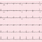
UMMC Cases Part III: When T Waves Help Tell the Story
A 23-year-old woman presents with palpitations and lightheadedness after a spontaneous episode of regular wide complex tachycardia. Her post-conversion ECG shows anterior T wave inversions with a subtle abnormality hiding…

Hypokalemia
Key Points: Hypokalemia delays ventricular repolarization, producing progressive changes in the T-U complex before serious arrhythmias develop. ECG progression: T-wave flattening → mild ST depression → prominent U waves →…
Hypokalemia Emergencies
Key Points: Severe hypokalemia can result in an arrhythmia emergency. The major threat is ventricular tachyarrhythmias, including torsades de pointes, polymorphic ventricular tachycardia, ventricular fibrillation, and sudden cardiac death. Severe…
Hypermagnesemia
Key Points: Hypermagnesemia is uncommon and usually occurs when impaired renal magnesium clearance is combined with increased magnesium exposure, such as magnesium-containing laxatives, antacids, bowel preparations, or therapeutic magnesium administration….
Hypomagnesemia
Key Points: Hypomagnesemia is an important arrhythmogenic electrolyte abnormality. It can promote atrial and ventricular ectopy and increase susceptibility to ventricular arrhythmias, particularly when other repolarization abnormalities are present. ECG…
Hypocalcemia
Key Points: ECG hallmark: QT prolongation caused predominantly by ST-segment prolongation, usually with relatively preserved T-wave morphology. Arrhythmia risk: Severe hypocalcemia can predispose to ventricular arrhythmias, including torsades de pointes…
Severe Hypothermia
Key Points: Severe hypothermia produces progressive electrical slowing. Sinus bradycardia, PR prolongation, QRS widening, QT prolongation, and atrial fibrillation with a slow ventricular response become increasingly common as core temperature…
Ventricular Tachycardia (VT) Mimics
Key Points: Default assumption: A regular wide-complex tachycardia should be considered VT until another diagnosis is more likely. Rate modifies the differential, not the diagnosis. Typical monomorphic VT is often…
Hyperkalemic Periodic Paralysis (Impressive Syndrome)
Key Points: What It Is: Severe hyperkalemia can cause sudden, profound, symmetric muscle weakness or flaccid paralysis. Dr. Mattu has referred to this memorable presentation as “Impressive Syndrome,” a teaching…
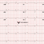
The Cath Lab Was Activated, But Something Didn’t Fit
A 70-year-old woman with CHF, COPD, intermittent atrial fibrillation, chronic pain medication use, and recent poor intake develops sudden dyspnea at rest and is found somnolent and bradycardic in the…
Hyperkalemia Emergencies
Key Points: Severe Hyperkalemia Mimics Several Life-Threatening Conditions: Severe hyperkalemia is one of the most dangerous ECG mimics in emergency medicine. It can resemble unstable bradyarrhythmias, VT, STEMI, and pacemaker…
Short QT Interval: DDx
Key Points: Short QT Interval: A QT interval is considered short when the corrected QT (QTc) interval is less than 350 ms. A short QT interval on the ECG can…
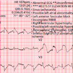
Baltimore City EMS ECGs: Pitfalls and Mimics (Part 2)
A 54-year-old man presents to the emergency department by EMS with acute shortness of breath. A prehospital ECG triggers a STEMI alert based on the computer interpretation. The tracing shows…
Hypercalcemia
Key Points: ECG hallmark: Shortened QT interval, primarily due to shortening of the ST segment. STEMI mimic: Severe hypercalcemia can produce apparent ST-segment elevation and a pseudoinfarction pattern, occasionally mimicking…
Hypokalemic Periodic Paralysis
Key Points: Definition: Hypokalemic periodic paralysis (HPP) is a rare disorder characterized by episodic, reversible flaccid muscle weakness associated with a shift of potassium from the extracellular space into skeletal…
STEMI vs Severe Hyperkalemia: DDx
Key Points: Severe hyperkalemia is a true ECG chameleon. It can produce ST elevation, wide QRS complexes, axis shifts, and conduction blocks that closely mimic STEMI or ventricular tachycardia. New…
Himalayan T Waves
Key Points Tall, broad-based T–U fusion that looks like a mountain peak, usually from severe hypokalemia; think high torsades risk until proven otherwise. Hallmark is prolonged repolarization: QT appears long…
Osborn (J) Wave: Basics
Key Points An Osborn wave is a notch or slur at the J point that becomes more prominent as core temperature falls. Most visible in inferolateral and precordial leads; can…
BRASH Syndrome
Key Points BRASH is a synergistic spiral: bradycardia, renal failure, therapeutic AV-nodal blockade, shock, and hyperkalemia. The signature clue is disproportionate brady-shock despite only modest potassium elevation. Do not be…
U Wave: Basics
Key Points Definition and origin: The U wave is a small deflection following the T wave, best seen in V2–V3. It likely reflects late ventricular repolarization or Purkinje repolarization. Normal…
ECG Tags
- 5th Universal Definition of MI
- A-Z
- ACS Mimics
- ACS-OMI
- Activation
- Acute MI
- Advanced Level Curriculum
- Annual ECG Competition
- Anterior OMI
- Anterior STEMI
- Approach
- Arrest
- Arrhythmia
- Arrhythmogenic Cardiomyopathy
- Artifact
- ARVC
- ARVD
- Atrial Parasystole
- Attending
- AV Block
- aVF
- aVL
- aVR
- Axis
- Barcelona
- Basics
- Bix Rule
- Board Review
- Bradyarrhythmia
- Cath Lab
- Chest Pain
- Clumped Beats
- Competition
- Conduction
- Core
- Core Level Curriculum
- Critical ECG Patterns
- Curriculum
- DDx
- Delta
- Devices
- Diagonal Branch Occlusion
- Differential Diagnoses
- Diffuse ST Elevation
- Documentation
- Early Repolarization
- ECG Interpretation
- ECG Localization
- ECG Variant
- Education/Teaching
- Electrolytes
- Emergencies
- Emergent Cath Lab Activation
- EMS
- Expert Level Curriculum
- Flutter
- Flutter Waves
- Foundations Level Curriculum
- Guidelines
- High-Lateral STEMI
- Hub
- Hyperacute T waves
- Hypercalcemia
- Hyperkalemia
- Hypermagnesemia
- Hypocalcemia
- Hypokalemia
- Hypomagnesemia
- Hyponatremia
- Hypothermia
- I
- II
- Index
- Inferior OMI
- Inferior STEMI
- Intervals
- Irregular
- Ischemia
- Ischemia & Infarction
- J point
- J Waves
- JT
- Junctional Rhythm
- Juvenile T wave
- LAD Occlusion
- Lateral STEMI
- LBBB
- LCx Occlusion
- Lead Placement
- Left Atrial Enlargement (LAE)
- Life Savers
- LV an
- LV Aneurysm
- LVAD
- Mastery Level Curriculum
- Metabolic
- MI
- Mimics
- Morphology
- Narrow QRS
- Occlusion MI
- OMI Pattern
- Orthodromic AVRT
- Osborn Waves
- P Wave
- Paced Rhythms
- Pacemaker/ICD
- Paramedics
- Pauses
- PE
- Pediatrics
- Pericardial
- Pericarditis
- PGY-1
- PGY-2
- PGY-3
- PGY-4
- Post-Arrest
- post-arrest STEMI
- Post-Cardiac Arrest
- Posterior Extension
- Posterior MI
- Posterior STEMI
- potpourri
- PR
- Preexcitation
- Premature Complexes
- Prolonged QT
- Pulmonary Embolism
- Pulse-Tapping Artifact
- PVCs
- Q Wave
- QRS
- QT
- R Waves
- RAD
- Rate
- RBBB
- RCA Occlusion
- Regular
- Reperfusion
- Rhythm
- Right Atrial Enlargement (RAE)
- RR
- S Waves
- S1Q3T3
- Segments
- Seizure
- Serial ECGs
- Sgarbossa
- Shock
- South African Flag Sign
- ST
- ST Depression
- ST Elevation
- STEMI
- STEMI Equivalent
- STEMI Mimics
- STEMI Negative OMI
- Stepwise
- Stroke
- Structural
- Students
- SVT
- Syncope
- T Wave Inversion
- T Waves
- Tachyarrhythmia
- Tachycardia
- Tamponade
- Terminal QRS Distortion
- Toxicology
- TP
- Traditional STEMI Criteria
- U Waves
- Unstable
- V1-V4
- V2
- V5
- V6
- Vectors
- Ventricular Repolarization
- Ventricular Rhythms
- Ventricular Tachycardia
- Voltage
- Waveforms
- Wide QRS
- Workflow
- WPW
© 2026 ECG Weekly. All rights reserved. | Terms of Use | Privacy Policy
Powered by the member(dev) platform
 Loading...