Conduction
Results:
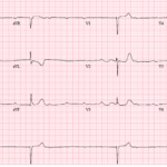
The Syncope ECG With Too Much P
A 68-year-old man has syncope, then has a second syncopal episode while lying still on a stretcher during evaluation at an outpatient clinic. He is sent emergently to the ED….

Isorhythmic AV Dissociation
Key Points: Isorhythmic AV dissociation is a form of AV dissociation in which the sinus rate and junctional or ventricular escape rate are nearly identical, making the P waves and…
Third-Degree AV Block (Complete Heart Block)
Key Points: Third-degree AV block is complete failure of atrial impulses to conduct to the ventricles. The defining ECG feature is AV dissociation with no conducted P waves. The atrial…
Second-Degree AV Block Type I (Mobitz I/Wenckebach)
Key Points: Mobitz I is defined by progressive PR prolongation until a single P wave fails to conduct, after which the cycle resets. The block is usually at the AV…
Second-Degree AV Block with 2:1 Conduction
Key Points: Second-degree AV block with 2:1 conduction means every other P wave conducts and every other P wave is blocked. A single ECG with 2:1 conduction usually cannot be…
First-Degree AV Block
Key Points: First-degree AV block is defined by a PR interval greater than 200 ms with fixed 1:1 AV conduction and no dropped QRS complexes. It usually reflects delayed conduction,…
Second-Degree AV Block Type II (Mobitz II)
Key Points: Mobitz II is defined by sudden failure of AV conduction after at least 2 consecutive conducted beats with fixed PR intervals and no preceding PR prolongation. The block…
Atrial Flutter
Key Points: Atrial flutter is a supraventricular tachyarrhythmia caused by a macro-reentrant circuit, most commonly typical cavotricuspid isthmus-dependent flutter in the right atrium. The atrial rate is usually about 250-350…
Advanced (High-Grade) AV Block
Key Points: Advanced or high-grade AV block is a severe second-degree AV block with 2 or more consecutive non-conducted P waves, such as 3:1 or 4:1 conduction. Do not force…
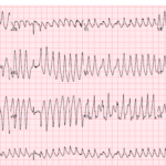
Preexcitation Pitfalls (Part 3): Wide, Irregular, Fast…Avoid AV Nodal Blockers
A 53-year-old man presents with palpitations and lightheadedness. The following ECG is obtained on arrival and appears very rapid and irregular with changing QRS morphologies. He starts showing signs of…
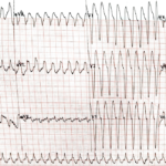
Preexcitation Pitfalls (Part 2): Wide, Regular, Fast…Treat It Like VT
A young man with recurrent palpitations presents to the emergency department hemodynamically stable during an episode. The arrival ECG shows a wide complex, regular tachycardia and the computer interpretation calls…
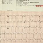
Preexcitation Pitfalls (Part 1): The “Inferior STEMI” That Isn’t
A critically ill 38-year-old man presents hypotensive, pale, and diaphoretic with abdominal pain and rectal bleeding. Upright chest X-ray shows free air under the diaphragm, and the patient is headed…
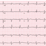
ECG Foundations: Vectors, Leads, & Activation
Key Points: An ECG records voltage differences over time. The ECG tracing is a plot where the horizontal axis is time and the vertical axis is voltage. Leads are viewpoints….
Dr. Mattu’s 5 Favorite ECG Cases of 2025
A 68-year-old man presents after syncope with profound bradycardia. The ECG shows a very slow ventricular rate with high-grade AV block. The reflex move is to focus only on pacing,…
Premature Atrial Complexes (PACs)
Key Points PACs are early atrial depolarizations from an ectopic focus that create a premature P wave with a different morphology and axis than the sinus P wave, usually followed…
Premature Junctional Complexes (PJCs)
Key Points PJCs are premature impulses from ectopic foci in or near the AV junction. ECG hallmark is a narrow premature beat with an absent or retrograde P wave. Retrograde…
Blocked Premature Atrial Complexes (PACs)
Key Points Definition: early ectopic atrial beats that do not conduct to the ventricles. You see a premature P wave with no following QRS and a pause that is usually…
Premature Complexes (PACs, PJCs, & PVCs) Overview
Key Points: Premature complexes are early depolarizations arising from the atrium, AV junction, or ventricle which interrupt the expected sinus rhythm. Rapid classification by origin: look for a P wave…
Premature Ventricular Complexes (PVCs)
Key Points PVCs are early ventricular depolarizations that produce a wide QRS with secondary ST-T changes and are usually followed by a full compensatory pause. No preceding P wave. A…
Fascicular Blocks
Key Points Definition: Delay/block within the left bundle’s fascicles—left anterior (LAF) or left posterior (LPF)—alters ventricular activation and the QRS axis. Types: LAFB (common) → left axis deviation; LPFB (rare)…
ECG Tags
- A-Z
- ACS Mimics
- ACS-OMI
- Activation
- Advanced Level Curriculum
- Annual ECG Competition
- Anterior OMI
- Anterior STEMI
- Approach
- Arrest
- Arrhythmia
- Arrhythmogenic Cardiomyopathy
- Artifact
- ARVC
- ARVD
- Atrial Parasystole
- Attending
- AV Block
- aVF
- aVL
- aVR
- Axis
- Barcelona
- Basics
- Bix Rule
- Board Review
- Bradyarrhythmia
- Cath Lab
- Chest Pain
- Clumped Beats
- Competition
- Conduction
- Core
- Core Level Curriculum
- Critical ECG Patterns
- Curriculum
- DDx
- Delta
- Devices
- Diagonal Branch Occlusion
- Differential Diagnoses
- Diffuse ST Elevation
- Documentation
- Early Repolarization
- ECG Interpretation
- ECG Localization
- ECG Variant
- Education/Teaching
- Electrolytes
- Emergencies
- Emergent Cath Lab Activation
- EMS
- Expert Level Curriculum
- Flutter
- Flutter Waves
- Foundations Level Curriculum
- Guidelines
- High-Lateral STEMI
- Hub
- Hyperacute T waves
- Hypercalcemia
- Hyperkalemia
- Hypermagnesemia
- Hypocalcemia
- Hypokalemia
- Hypomagnesemia
- Hyponatremia
- Hypothermia
- I
- II
- Index
- Inferior OMI
- Inferior STEMI
- Intervals
- Irregular
- Ischemia
- Ischemia & Infarction
- J point
- J Waves
- JT
- Junctional Rhythm
- Juvenile T wave
- LAD Occlusion
- Lateral STEMI
- LBBB
- LCx Occlusion
- Lead Placement
- Left Atrial Enlargement (LAE)
- Life Savers
- LV an
- LV Aneurysm
- LVAD
- Mastery Level Curriculum
- Metabolic
- Mimics
- Morphology
- Narrow QRS
- Occlusion MI
- OMI Pattern
- Orthodromic AVRT
- Osborn Waves
- P Wave
- Paced Rhythms
- Pacemaker/ICD
- Paramedics
- Pauses
- PE
- Pediatrics
- Pericardial
- Pericarditis
- PGY-1
- PGY-2
- PGY-3
- PGY-4
- Post-Arrest
- post-arrest STEMI
- Post-Cardiac Arrest
- Posterior Extension
- Posterior MI
- Posterior STEMI
- potpourri
- PR
- Preexcitation
- Premature Complexes
- Prolonged QT
- Pulmonary Embolism
- Pulse-Tapping Artifact
- PVCs
- Q Wave
- QRS
- QT
- R Waves
- RAD
- Rate
- RBBB
- RCA Occlusion
- Regular
- Reperfusion
- Rhythm
- Right Atrial Enlargement (RAE)
- RR
- S Waves
- S1Q3T3
- Segments
- Seizure
- Serial ECGs
- Sgarbossa
- Shock
- South African Flag Sign
- ST
- ST Depression
- ST Elevation
- STEMI
- STEMI Equivalent
- STEMI Mimics
- STEMI Negative OMI
- Stepwise
- Stroke
- Structural
- Students
- SVT
- Syncope
- T Wave Inversion
- T Waves
- Tachyarrhythmia
- Tachycardia
- Tamponade
- Terminal QRS Distortion
- Toxicology
- TP
- Traditional STEMI Criteria
- U Waves
- Unstable
- V1-V4
- V2
- V5
- V6
- Vectors
- Ventricular Repolarization
- Ventricular Rhythms
- Ventricular Tachycardia
- Voltage
- Waveforms
- Wide QRS
- Workflow
- WPW
© 2026 ECG Weekly. All rights reserved. | Terms of Use | Privacy Policy
Powered by the member(dev) platform
 Loading...