Approach
Results:
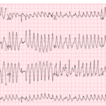
Preexcitation Pitfalls (Part 3): Wide, Irregular, Fast…Avoid AV Nodal Blockers
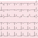
A 53-year-old man presents with palpitations and lightheadedness. The following ECG is obtained on arrival and appears very rapid and irregular with changing QRS morphologies. He starts showing signs of…
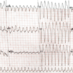
Preexcitation Pitfalls (Part 2): Wide, Regular, Fast…Treat It Like VT
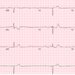
A young man with recurrent palpitations presents to the emergency department hemodynamically stable during an episode. The arrival ECG shows a wide complex, regular tachycardia and the computer interpretation calls…

Appropriate Discordance
Key Points: Appropriate discordance refers to the expected secondary ST segment and T wave pattern seen with abnormal ventricular depolarization, especially LBBB and ventricular-paced rhythm. The ST segment and T…
QRS Morphology and ST-T Interpretation: Basics
Key Points: Read the QRS before you read the ST segment or T wave. Ventricular depolarization shapes repolarization. Narrow QRS usually reflects normal His-Purkinje conduction. Wide QRS suggests abnormal ventricular…
Preexcitation Pitfalls (Part 1): The “Inferior STEMI” That Isn’t
A critically ill 38-year-old man presents hypotensive, pale, and diaphoretic with abdominal pain and rectal bleeding. Upright chest X-ray shows free air under the diaphragm, and the patient is headed…

Stepwise Approach to STAT ECGs Hub
Key Points: Consistency saves lives: Use a repeatable ECG routine to reduce misses in chaotic settings. Many valid methods exist. Pick an order that fits your acute-care workflow and do…
Three More ECG Pitfalls That Punish Anchoring Bias
A 51-year-old man with lung cancer presents with shortness of breath and tachycardia. The arrival ECG shows an S1Q3 pattern and seems to support a familiar diagnosis that would normally…
Four ECG Pitfalls That Punish Anchoring Bias
A 43-year-old woman with sharp left-sided chest pain and minimal cardiac risk factors has an initial ECG that is not diagnostic for STEMI. She looks stable, but one feature on…
Waveforms, Segments, & Intervals: Basics
Key Points: Every ECG tracing is built from waveforms (deflections), segments (baseline portions between waveforms), and intervals (time that include waveforms plus segments). Waveforms describe electrical events (depolarization or repolarization)….
STAT ECG 101: The Acute Care ECG Workflow
Key Points: The ECG is the fastest bedside test for rhythm, conduction, ischemia, and tox-metabolic disease. It only saves lives when interpreted systematically and acted on. In acute care, the…
Dr. Mattu’s 5 Favorite ECG Cases of 2025
A 68-year-old man presents after syncope with profound bradycardia. The ECG shows a very slow ventricular rate with high-grade AV block. The reflex move is to focus only on pacing,…
J Point: Basics
Key Points The J point is the junction where QRS ends and the ST segment begins. It is a location, not a waveform. ST deviation is judged at the J…
R Wave: Basics
Key Points Definition: The R wave is the first positive deflection of the QRS complex, reflecting early ventricular depolarization, predominantly of the left ventricle. Normal progression: Precordial R amplitude increases…
Wide & Regular Tachydysrhythmia DDx
Key Points Definition: Wide complex tachycardia (WCT) = QRS >120 ms with a steady R-R interval. This section focuses on regular WCT (RWCT). Wide & irregular rhythms are covered separately…
Wide QRS Complex: DDx
Key Points: A QRS duration greater than 120 ms indicates abnormal ventricular depolarization. A wide QRS can signal conditions that range from benign to immediately life-threatening. Developing a focused differential…
Ischemia & Infarction Interpretation
Key Points: The ECG is the fastest bedside tool for detecting acute coronary occlusion and dynamic ischemia, often before troponin changes and sometimes before classic symptoms. Acute coronary syndromes are…
Irregularly Irregular Rhythm: Basics
Key Points: Definition: An irregularly irregular rhythm occurs when the R-R intervals or P-P intervals vary with no consistent pattern, making the rhythm unpredictable and abnormal. Clinical Significance: Identifying an…
Heart Rhythm: Basics
Key Points Don’t trust the ECG machines automated interpretation. Confirm the rhythm yourself. Start with the ventricles (R–R pattern), then the atria (P waves), then the AV relationship (PR behavior/P:QRS)….
Heart Rate: Basics
Key Points: Never accept the machine’s rate blindly. Confirm it yourself as ECG computer interpretations are frequently inaccurate. Verify paper speed and gain first (default 25 mm/s, 10 mm/mV). Name…
Heart Rate & Rhythm Interpretation
Key Points: Treat the patient, not just the number or rhythm. Start with a 10-second stability check. If the rhythm explains hypotension, shock, ischemic chest pain, altered mentation, or severe…
ECG Tags
- A-Z
- ACS Mimics
- ACS-OMI
- Activation
- Advanced Level Curriculum
- Annual ECG Competition
- Anterior OMI
- Anterior STEMI
- Approach
- Arrest
- Arrhythmia
- Arrhythmogenic Cardiomyopathy
- Artifact
- ARVC
- ARVD
- Atrial Parasystole
- Attending
- AV Block
- aVF
- aVL
- aVR
- Axis
- Basics
- Board Review
- Bradyarrhythmia
- Chest Pain
- Clumped Beats
- Conduction
- Core
- Core Level Curriculum
- Critical ECG Patterns
- Curriculum
- DDx
- Delta
- Devices
- Diagonal Branch Occlusion
- Differential Diagnoses
- Diffuse ST Elevation
- Documentation
- Early Repolarization
- ECG Interpretation
- ECG Localization
- ECG Variant
- Education/Teaching
- Electrolytes
- Emergencies
- Emergent Cath Lab Activation
- EMS
- Expert Level Curriculum
- Foundations Level Curriculum
- Guidelines
- High-Lateral STEMI
- Hub
- Hyperacute T waves
- Hypercalcemia
- Hyperkalemia
- Hypermagnesemia
- Hypocalcemia
- Hypokalemia
- Hypomagnesemia
- Hyponatremia
- Hypothermia
- I
- II
- Index
- Inferior OMI
- Inferior STEMI
- Intervals
- Irregular
- Ischemia
- Ischemia & Infarction
- J Waves
- JT
- Juvenile T wave
- LAD Occlusion
- Lateral STEMI
- LBBB
- LCx Occlusion
- Lead Placement
- Life Savers
- LV an
- LV Aneurysm
- Mastery Level Curriculum
- Metabolic
- Mimics
- Morphology
- Narrow QRS
- Occlusion MI
- OMI Pattern
- Orthodromic AVRT
- Osborn Waves
- P Wave
- Paced Rhythms
- Pacemaker/ICD
- Paramedics
- Pauses
- PE
- Pediatrics
- Pericardial
- Pericarditis
- PGY-1
- PGY-2
- PGY-3
- PGY-4
- Post-Cardiac Arrest
- Posterior Extension
- Posterior MI
- Posterior STEMI
- PR
- Preexcitation
- Premature Complexes
- Prolonged QT
- Pulmonary Embolism
- Pulse-Tapping Artifact
- PVCs
- Q Wave
- QRS
- QT
- R Waves
- RAD
- Rate
- RBBB
- RCA Occlusion
- Regular
- Reperfusion
- Rhythm
- RR
- S Waves
- Segments
- Seizure
- Serial ECGs
- Sgarbossa
- Shock
- ST
- ST Depression
- ST Elevation
- STEMI
- STEMI Equivalent
- STEMI Mimics
- STEMI Negative OMI
- Stepwise
- Stroke
- Structural
- Students
- SVT
- Syncope
- T Wave Inversion
- T Waves
- Tachyarrhythmia
- Tachycardia
- Tamponade
- Terminal QRS Distortion
- Toxicology
- TP
- Traditional STEMI Criteria
- U Waves
- Unstable
- V1-V4
- V5
- V6
- Vectors
- Ventricular Repolarization
- Ventricular Rhythms
- Ventricular Tachycardia
- Voltage
- Waveforms
- Wide QRS
- Workflow
- WPW
© 2026 ECG Weekly. All rights reserved. | Terms of Use | Privacy Policy
Powered by the member(dev) platform
 Loading...