Mimics
Results:

Wide QRS Complex: DDx
Key Points: A QRS duration greater than 120 ms reflects delayed or abnormal ventricular depolarization. A wide QRS may be chronic and benign in context, or it may be the…
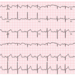
UMEM Cases, Part 3: When the Diagnosis Seems Clear and When It Is Not
A 71-year-old man presents with shortness of breath, and his ECG is initially read as a junctional rhythm. On later review, it is even mistaken for atrial fibrillation. But the…
ST Elevation in aVR: DDx
Key Points: ST elevation (STE) in aVR with diffuse ST depression most often reflects global subendocardial ischemia, not focal transmural infarction. High-risk coronary disease is one cause, not the only…
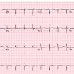
UMEM Cases, Part 1: When the ECG Lies and When It Evolves
A 72-year-old man presents with chest pain and shortness of breath. His ECG shows sinus rhythm with LVH, mild inferior ST elevation, and lateral ST-T abnormalities that some interpret as…
STEMI vs LVH: DDx
Key Points: Left ventricular hypertrophy (LVH) with strain is one of the most common and dangerous STEMI mimics, particularly in the anterior leads, and is a frequent cause of false-positive…
WPW Syndrome and Pseudo-MI Patterns
Key Points: WPW alters ventricular depolarization, producing secondary repolarization abnormalities that can mimic or mask myocardial infarction. ST-segment deviation in WPW is often non-ischemic, driven by abnormal activation via the…
ST Elevation: DDx
Key Points: ST elevation describes an ECG finding, not a diagnosis. It reflects abnormal ventricular repolarization and can arise from ischemic, structural, metabolic, electrical, or extracardiac processes. Occlusion MI is…
STEMI vs LV Aneurysm: DDx
Key Points: LV aneurysm pattern is a post MI scar pattern with persistent ST elevation in the prior infarct territory, usually with pathologic Q waves and a stable, non evolving…
STEMI Mimics: DDx
Key Points: ST elevation is a pattern, not a diagnosis. STEMI represents one cause of ST elevation and requires correlation with ECG morphology, distribution, evolution, and clinical context. Most ED…
STEMI vs Takotsubo Cardiomyopathy: DDx
Key Points: Takotsubo (stress) cardiomyopathy is a transient, non-ischemic LV dysfunction—classically apical ballooning with basal hyperkinesis—often after emotional or physical stress. Presentation mimics occlusion MI (chest pain, ECG changes, elevated…
Pulse-Tapping Artifact
Key Points: Mechanical artifact caused by an ECG electrode sitting on top of a strong arterial pulse. Seen frequently in dialysis patients with AV fistulas. Can mimic serious pathology including…
Ventricular Tachycardia (VT) Mimics
Key Points: Initial Assumption: Any wide (QRS >120 ms), regular tachycardia should be considered ventricular tachycardia (VT) until clearly proven otherwise. VT Characteristics: VT generally has a ventricular rate of…
Wide & Regular Tachydysrhythmia DDx
Key Points: Definition: Wide complex tachycardia (WCT) = QRS >120 ms with a steady R-R interval. This section focuses on regular WCT (RWCT). Wide & irregular rhythms are covered separately…
VT vs SVT with Aberrancy: DDx
Key Points ECG alone cannot reliably distinguish VT from SVT-aberrancy in many cases. Use ECG features to rule in VT, not to exclude it. Default: treat regular WCT as VT…
Understanding ECG Artifacts
Key Points Artifacts = non-cardiac signals that distort or obscure true ECG. They come from the patient, leads/equipment, or the environment. Clues to artifact: lacks a physiologic pattern, varies beat-to-beat…
Slow Ventricular Tachycardia (VT)
Key Points Definition: Slow ventricular tachycardia is defined as a wide complex tachycardia with a ventricular rate between 100–120 bpm. Distinction: Typical sustained VT usually exceeds 120 bpm. When encountering…
Terminal QRS Distortion: OMI Pattern
Key Points: What it is: In V2 or V3, there is no S wave (the R does not descend below the PQ baseline) and no J wave (no notch/slur at…

Shock first and ask questions later?
An elderly woman with a past medical history of hypertension, diabetes, and Parkinson’s disease presents to the ED with hip pain after a fall. Her blood pressure and mental status…
ECG Tags
- A-Z
- ACS Mimics
- ACS-OMI
- Activation
- Advanced Level Curriculum
- Annual ECG Competition
- Anterior OMI
- Anterior STEMI
- Approach
- Arrest
- Arrhythmia
- Arrhythmogenic Cardiomyopathy
- Artifact
- ARVC
- ARVD
- Atrial Parasystole
- Attending
- AV Block
- aVF
- aVL
- aVR
- Axis
- Barcelona
- Basics
- Bix Rule
- Board Review
- Bradyarrhythmia
- Cath Lab
- Chest Pain
- Clumped Beats
- Competition
- Conduction
- Core
- Core Level Curriculum
- Critical ECG Patterns
- Curriculum
- DDx
- Delta
- Devices
- Diagonal Branch Occlusion
- Differential Diagnoses
- Diffuse ST Elevation
- Documentation
- Early Repolarization
- ECG Interpretation
- ECG Localization
- ECG Variant
- Education/Teaching
- Electrolytes
- Emergencies
- Emergent Cath Lab Activation
- EMS
- Expert Level Curriculum
- Flutter
- Flutter Waves
- Foundations Level Curriculum
- Guidelines
- High-Lateral STEMI
- Hub
- Hyperacute T waves
- Hypercalcemia
- Hyperkalemia
- Hypermagnesemia
- Hypocalcemia
- Hypokalemia
- Hypomagnesemia
- Hyponatremia
- Hypothermia
- I
- II
- Index
- Inferior OMI
- Inferior STEMI
- Intervals
- Irregular
- Ischemia
- Ischemia & Infarction
- J Waves
- JT
- Junctional Rhythm
- Juvenile T wave
- LAD Occlusion
- Lateral STEMI
- LBBB
- LCx Occlusion
- Lead Placement
- Left Atrial Enlargement (LAE)
- Life Savers
- LV an
- LV Aneurysm
- LVAD
- Mastery Level Curriculum
- Metabolic
- Mimics
- Morphology
- Narrow QRS
- Occlusion MI
- OMI Pattern
- Orthodromic AVRT
- Osborn Waves
- P Wave
- Paced Rhythms
- Pacemaker/ICD
- Paramedics
- Pauses
- PE
- Pediatrics
- Pericardial
- Pericarditis
- PGY-1
- PGY-2
- PGY-3
- PGY-4
- Post-Arrest
- post-arrest STEMI
- Post-Cardiac Arrest
- Posterior Extension
- Posterior MI
- Posterior STEMI
- potpourri
- PR
- Preexcitation
- Premature Complexes
- Prolonged QT
- Pulmonary Embolism
- Pulse-Tapping Artifact
- PVCs
- Q Wave
- QRS
- QT
- R Waves
- RAD
- Rate
- RBBB
- RCA Occlusion
- Regular
- Reperfusion
- Rhythm
- Right Atrial Enlargement (RAE)
- RR
- S Waves
- Segments
- Seizure
- Serial ECGs
- Sgarbossa
- Shock
- South African Flag Sign
- ST
- ST Depression
- ST Elevation
- STEMI
- STEMI Equivalent
- STEMI Mimics
- STEMI Negative OMI
- Stepwise
- Stroke
- Structural
- Students
- SVT
- Syncope
- T Wave Inversion
- T Waves
- Tachyarrhythmia
- Tachycardia
- Tamponade
- Terminal QRS Distortion
- Toxicology
- TP
- Traditional STEMI Criteria
- U Waves
- Unstable
- V1-V4
- V2
- V5
- V6
- Vectors
- Ventricular Repolarization
- Ventricular Rhythms
- Ventricular Tachycardia
- Voltage
- Waveforms
- Wide QRS
- Workflow
- WPW
© 2026 ECG Weekly. All rights reserved. | Terms of Use | Privacy Policy
Powered by the member(dev) platform
 Loading...