Emergencies
Results:

WPW with Orthodromic SVT (Orthodromic AVRT)
Key Points: Orthodromic AVRT is the most common tachyarrhythmia in WPW and presents as a regular narrow-complex SVT that is indistinguishable from AVNRT during the tachycardia. Mechanism: antegrade conduction down…
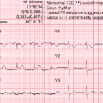
Baltimore City EMS ECGs: Pitfalls and Mimics (Part 2)
A 54-year-old man presents to the emergency department by EMS with acute shortness of breath. A prehospital ECG triggers a STEMI alert based on the computer interpretation. The tracing shows…
Third-Degree AV Block (Complete Heart Block)
Key Points: Definition: Third-degree AV block is complete failure of conduction from atria to ventricles, resulting in independent atrial and ventricular activity—known as AV dissociation. Hallmark Feature: No P waves…
Second-Degree AV Block Type I (Mobitz I/Wenckebach)
Key Points: Definition: Progressive PR interval prolongation until one atrial impulse fails to conduct to the ventricles (P wave is non-conducted), after which the cycle repeats. Site of Block: Typically…
Second-Degree AV Block with 2:1 Conduction
Key Points: Definition: A form of second-degree AV block in which every other atrial impulse is blocked, producing a 2:1 atrioventricular conduction ratio. Typing Limitation: Differentiating between Mobitz I and…
High-Grade (Advanced) AV Block
Key Points: Definition: A severe form of second-degree AV block with two or more consecutive non‑conducted P waves (for example 3:1, 4:1). Do not force a Mobitz label when multiple…
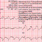
Baltimore City EMS ECGs: Pitfalls and Mimics (Part 1)
A 68-year-old man is brought to the emergency department by EMS with acute chest discomfort. The following prehospital ECG was obtained and shows concave ST elevation across multiple leads. The…
Three More ECG Pitfalls That Punish Anchoring Bias
A 51-year-old man with lung cancer presents with shortness of breath and tachycardia. The arrival ECG shows an S1Q3 pattern and seems to support a familiar diagnosis that would normally…
Bidirectional Ventricular Tachycardia (BiVT)
Key Points: BiVT is a regular wide-complex tachycardia with strict beat-to-beat alternation of QRS axis and/or bundle-branch pattern (often an approximately 180° frontal-plane axis flip). In adults, assume digoxin toxicity…
Four ECG Pitfalls That Punish Anchoring Bias
A 43-year-old woman with sharp left-sided chest pain and minimal cardiac risk factors has an initial ECG that is not diagnostic for STEMI. She looks stable, but one feature on…
Occlusion MI: STEMI Criteria & Beyond
Key Points: The ECG’s primary role in ACS is detecting acute coronary occlusion. Acute coronary occlusion myocardial infarction (OMI) is a time-critical diagnosis that requires immediate reperfusion. Time is myocardium….
STEMI (-) Occlusion MI: OMI Patterns
Key Points: Traditional STEMI criteria miss many acute coronary occlusions. A substantial proportion of true OMIs do not meet classic millimetric STEMI thresholds. OMI is a pathophysiologic diagnosis, not an…
Prolonged QT Interval: DDx
Key Points: The QT interval reflects the time it takes for total ventricular depolarization and repolarization (Q wave onset to T wave end). QT prolongation increases the risk of torsades…
Hypocalcemia
Key Points: Prolonged QTc is the hallmark ECG change in hypocalcemia, driven mainly by ST-segment prolongation with relatively normal T-wave shape. Hypocalcemia can increase arrhythmia risk, including TdP, but TdP…
STEMI in the Presence of Baseline ECG Abnormalities
Key Points: Baseline ECG abnormalities do not protect patients from occlusion MI. They increase miss rates because they distort the ST segment and T waves. The core question is not…
Lateral and High-Lateral STEMI: Criteria, Localization, and Pitfalls
Key Points: Lateral and high-lateral STEMI often present with subtle ST elevation and are commonly missed or labeled as nonspecific ST-T changes. Small-appearing ECG changes may represent true coronary occlusion…
Inferior STEMI: Criteria, RV Involvement, and Pitfalls
Key Points: Inferior STEMI is the most common STEMI subtype and is frequently complicated by right ventricular and posterior involvement. Inferior occlusion may present with classic ST elevation, subtle ischemic…
Anterior STEMI: Criteria, Localization, and Pitfalls
Key Points: Anterior STEMI represents large myocardial territory at risk and carries the highest mortality among STEMI subtypes. Early recognition and reperfusion are critical. LAD occlusion may present with classic…
ECG Findings of LV Aneurysm
Key Points: Definition: A true LV aneurysm is a chronic, post transmural MI complication from scarred myocardium with akinetic or dyskinetic (paradoxical) wall motion. ECG hallmark: Persistent ST elevation in…
Posterior STEMI: Criteria & Pitfalls
Key Points: High risk of missed diagnosis. Isolated posterior occlusion MI is frequently missed because ST elevation is absent on the standard 12-lead ECG. Instead, posterior infarction most often presents…
ECG Tags
- A-Z
- ACS Mimics
- ACS-OMI
- Activation
- Advanced Level Curriculum
- Annual ECG Competition
- Anterior OMI
- Anterior STEMI
- Approach
- Arrest
- Arrhythmia
- Arrhythmogenic Cardiomyopathy
- Artifact
- ARVC
- ARVD
- Atrial Parasystole
- Attending
- AV Block
- aVF
- aVL
- aVR
- Axis
- Basics
- Board Review
- Bradyarrhythmia
- Chest Pain
- Clumped Beats
- Conduction
- Core
- Core Level Curriculum
- Critical ECG Patterns
- Curriculum
- DDx
- Delta
- Devices
- Diagonal Branch Occlusion
- Differential Diagnoses
- Diffuse ST Elevation
- Documentation
- Early Repolarization
- ECG Interpretation
- ECG Localization
- ECG Variant
- Education/Teaching
- Electrolytes
- Emergencies
- Emergent Cath Lab Activation
- EMS
- Expert Level Curriculum
- Foundations Level Curriculum
- Guidelines
- High-Lateral STEMI
- Hub
- Hyperacute T waves
- Hypercalcemia
- Hyperkalemia
- Hypermagnesemia
- Hypocalcemia
- Hypokalemia
- Hypomagnesemia
- Hyponatremia
- Hypothermia
- I
- II
- Index
- Inferior OMI
- Inferior STEMI
- Intervals
- Irregular
- Ischemia
- Ischemia & Infarction
- J Waves
- JT
- Juvenile T wave
- LAD Occlusion
- Lateral STEMI
- LBBB
- LCx Occlusion
- Lead Placement
- Life Savers
- LV an
- LV Aneurysm
- Mastery Level Curriculum
- Metabolic
- Mimics
- Morphology
- Narrow QRS
- Occlusion MI
- OMI Pattern
- Orthodromic AVRT
- Osborn Waves
- P Wave
- Paced Rhythms
- Pacemaker/ICD
- Paramedics
- Pauses
- PE
- Pediatrics
- Pericardial
- Pericarditis
- PGY-1
- PGY-2
- PGY-3
- PGY-4
- Post-Cardiac Arrest
- Posterior Extension
- Posterior MI
- Posterior STEMI
- PR
- Preexcitation
- Premature Complexes
- Prolonged QT
- Pulmonary Embolism
- Pulse-Tapping Artifact
- PVCs
- Q Wave
- QRS
- QT
- R Waves
- RAD
- Rate
- RBBB
- RCA Occlusion
- Regular
- Reperfusion
- Rhythm
- RR
- S Waves
- Segments
- Seizure
- Serial ECGs
- Sgarbossa
- Shock
- South African Flag Sign
- ST
- ST Depression
- ST Elevation
- STEMI
- STEMI Equivalent
- STEMI Mimics
- STEMI Negative OMI
- Stepwise
- Stroke
- Structural
- Students
- SVT
- Syncope
- T Wave Inversion
- T Waves
- Tachyarrhythmia
- Tachycardia
- Tamponade
- Terminal QRS Distortion
- Toxicology
- TP
- Traditional STEMI Criteria
- U Waves
- Unstable
- V1-V4
- V2
- V5
- V6
- Vectors
- Ventricular Repolarization
- Ventricular Rhythms
- Ventricular Tachycardia
- Voltage
- Waveforms
- Wide QRS
- Workflow
- WPW
© 2026 ECG Weekly. All rights reserved. | Terms of Use | Privacy Policy
Powered by the member(dev) platform
 Loading...